Campylobacter jejuni DNA PCR Quantitative Positive Control – Reference Material for Molecular Testing Precision

Campylobacter jejuni DNA PCR Quantitative Positive Control – High-Quality Reference Material for Molecular Detection
Meta Description: Learn about Campylobacter jejuni DNA PCR Quantitative Positive Controls, their structure, preparation, qPCR calibration role, and biosafety use in molecular research. Includes 20+ references to authoritative .edu and .gov sources.

Introduction: Why Campylobacter jejuni Controls Are Essential in Molecular Research

Campylobacter jejuni is a microaerophilic, spiral-shaped bacterium widely used in academic research for studying bacterial motility, genome plasticity, and environmental resilience.
In quantitative PCR (qPCR) or real-time PCR workflows, the Campylobacter jejuni DNA PCR Quantitative Positive Control serves as a standardized template for verifying assay sensitivity, reproducibility, and cross-run comparability.

Using a certified DNA control avoids variability between runs and helps laboratories confirm the linear dynamic range, amplification efficiency, and threshold cycle (Ct) reliability of their molecular assays.

Authoritative references such as the CDC Laboratory Guidance, NIH Genomic Research Portal, and USDA NAL Microbiology Guides describe how DNA reference materials stabilize assay validation frameworks across molecular platforms.
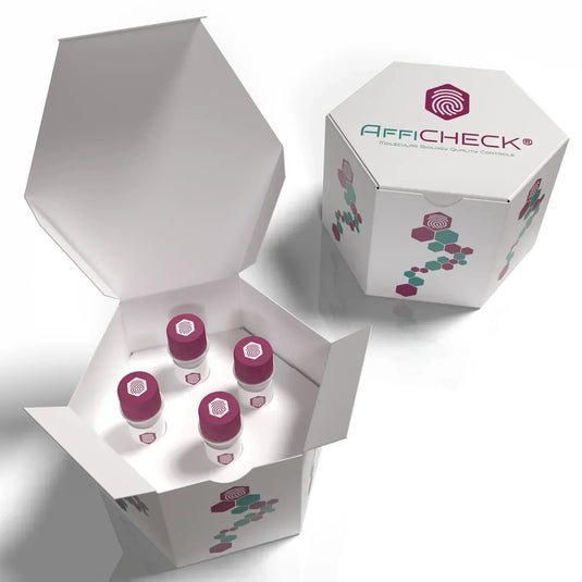
AffiCHECK® Campylobacter jejuni DNA PCR Quantitative Positive Control

Overview of Campylobacter jejuni DNA and Genome Structure

The Campylobacter jejuni genome is approximately 1.6–1.7 Mb with a G+C content near 30%. It encodes genes involved in:

  • Flagellar motility (critical for colonization and chemotaxis)

  • Iron acquisition and oxidative stress response

  • DNA methyltransferase systems contributing to phase variation

  • Metabolic adaptability for survival in microaerophilic environments

These genomic traits make C. jejuni an ideal control model in qPCR and RT-PCR for DNA integrity verification and amplification curve calibration.

For full genome references, consult the NCBI Genome Database, GenBank, and NCBI Bookshelf Microbiology Collection.

Role of Quantitative Positive Controls in PCR and qPCR Systems

A Quantitative Positive Control (QPC) is a DNA standard containing a known number of target copies per microliter.
When included in a PCR or qPCR assay, it allows researchers to:

  • Validate the analytical sensitivity of the amplification reagents

  • Monitor instrument calibration (e.g., light source intensity, detection thresholds)

  • Create standard curves for absolute quantification

  • Detect pipetting or reagent drift across runs

Typical concentrations range from 10³ to 10⁸ copies/µL, depending on the assay design.
These standards can be plasmid-based, linearized fragments, or synthetic gBlocks carrying a species-specific sequence, such as the mapA or hipO gene in C. jejuni.

For detailed principles of qPCR calibration and standards, see:

Campylobacter jejuni DNA Control Preparation and Composition

The Campylobacter jejuni DNA Quantitative Positive Control is produced by isolating high-purity genomic or plasmid DNA carrying a target-specific region.
Each batch typically undergoes:

  • Spectrophotometric analysis (A260/A280 ≥ 1.8)

  • Integrity testing by agarose gel electrophoresis

  • Concentration quantification by fluorometric assay (e.g., Qubit or PicoGreen)

  • Stability evaluation under long-term storage (−20 °C or below)

The DNA fragment corresponds to a unique region conserved within C. jejuni species, ensuring no cross-reactivity with C. coli, C. lari, or other Campylobacter spp.

For molecular biology standards, refer to NIST DNA Quantitation Standards and NIH Genetic Material Handling Guidelines.

Quantitative Curve Generation Using the Positive Control

Step-by-Step Standard Curve Setup:

  1. Prepare serial tenfold dilutions (e.g., 10⁸ → 10² copies/µL).

  2. Run each dilution in triplicate in the same plate.

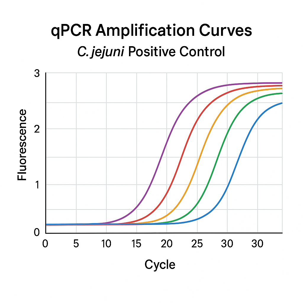
  3. Plot Ct values vs. log10(copy number) to determine slope and R².

  4. Calculate PCR efficiency (E) using the formula:

    E=10(−1/slope)−1E = 10^{(-1/slope)} – 1

    Acceptable range: 90–110%.

A well-calibrated standard curve ensures that unknown samples yield accurate quantification within the validated dynamic range.

More about amplification efficiency and standard curves can be found on:
Applications in Academic and Industrial Research

The Campylobacter jejuni DNA Positive Control supports various experimental goals:

  • Environmental microbiology: detecting Campylobacter persistence in water or food matrices.

  • Comparative genomics: calibrating inter-laboratory qPCR data.

  • Microbial ecology: tracking population abundance in mixed cultures.

  • Educational use: standard for teaching PCR technique reproducibility.

It is a non-hazardous reference standard that does not contain viable bacteria.
For biosafety, consult CDC BMBL and institutional biosafety guidelines such as Stanford Biosafety Manual or UC Berkeley Biosafety Manual.

Quality Control and Verification

To ensure precision, each batch is validated for:

  • Reproducibility: consistent Ct values across multiple runs.

  • Specificity: absence of amplification in non-target DNA.

  • Stability: minimal degradation after repeated freeze–thaw cycles.

  • Quantitative consistency: less than 5% variation across dilutions.

Data and reference certificates should accompany each lot, aligning with ISO 20395:2019 guidelines for quantitative molecular assays.

For reference material traceability, academic users often refer to NIST SRM Libraries and EPA Genomic Quality Control Resources.

Example Workflow Integration

A typical workflow for using this control includes:

  1. Reconstitute the DNA in nuclease-free water.

  2. Store aliquots at −20 °C to prevent degradation.

  3. Include 2–3 control concentrations in each qPCR run.

  4. Validate the limit of detection (LOD) using ≥20 replicates of low-concentration controls.

  5. Document and compare results between instruments or reagent lots.

This practice aligns with recommendations from NIH Reproducibility Initiatives and NIAID Standardization Guidelines.

Advantages of Using Certified Quantitative DNA Controls

  • Lot-to-lot consistency for longitudinal projects

  • Reduced variability across reagents and thermocyclers

  • Reliable amplification efficiency benchmarking

  • Improved publication reproducibility through standardized calibration

  • Safer handling—non-viable and ready-to-use DNA templates

Comprehensive laboratory practice is further supported by resources like the Harvard Recombinant DNA Policy and UCSF Biosafety Program.
Data Analysis and Interpretation Guidelines

During amplification:

  • The Ct value should decrease linearly with increasing copy number.

  • R² ≥ 0.99 indicates high precision.

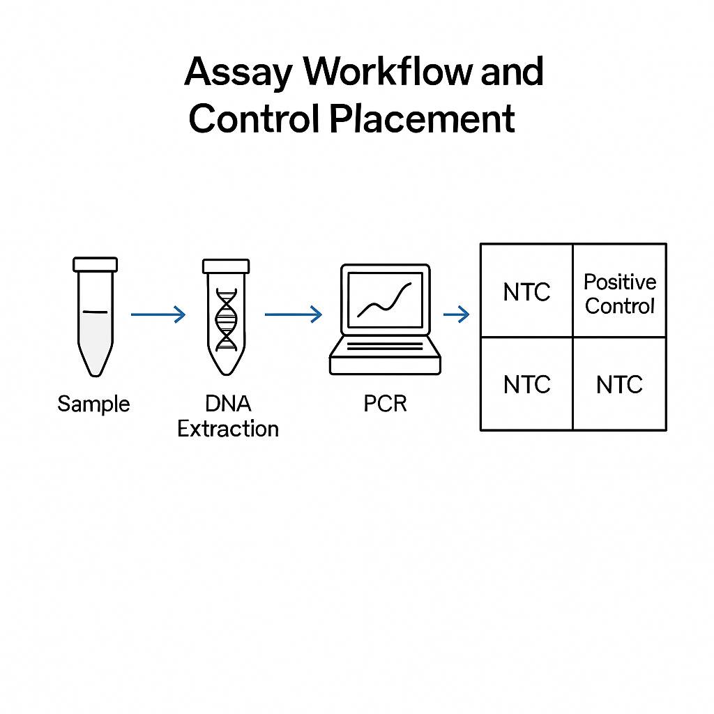
  • No-template controls (NTC) must show no amplification signal.

  • Efficiency deviations indicate pipetting or reagent inconsistencies.

For curve fitting and linear regression, NCBI Tools and NIH PMC Biostatistics Resources provide computational examples.

Storage and Stability Recommendations

The Campylobacter jejuni DNA Positive Control is stable for up to 24 months under proper storage.
Avoid multiple freeze–thaw cycles; divide the master stock into single-use aliquots.
Freeze at −20 °C or below and protect from repeated temperature fluctuations.

Stability testing follows standard molecular reagent handling practices per NIH Laboratory Best Practices and EPA DNA Integrity Guidelines.

Troubleshooting Tips

Observation Possible Cause Corrective Action
High Ct values DNA degradation or low template input Check concentration; use fresh aliquot
Non-linear curve Pipetting error or contamination Repeat dilutions under sterile conditions
No amplification Thermocycler calibration issue Verify annealing temperature and reagent quality
Variable triplicates Poor mixing Vortex thoroughly and briefly centrifuge before use

See related qPCR troubleshooting tutorials from NIH PMC and USDA Laboratory Guidebook.

Authoritative .edu / .gov References

  1. CDC Laboratory Reference Materials

  2. NIH Genomic Research Portal

  3. USDA ARS Genomics Program

  4. NCBI Genome Database

  5. NCBI Bookshelf Microbiology Collection

  6. NIST DNA Quantitation Standards

  7. NIH PMC qPCR Research

  8. FDA Genomics Resources

  9. EPA Genomic Quality Control

  10. Stanford Biosafety Manual

  11. UC Berkeley Biosafety Manual

  12. Harvard Recombinant DNA

The Campylobacter jejuni DNA PCR Quantitative Positive Control provides a reliable DNA standard for qPCR and PCR assay validation. It ensures consistent amplification performance, precise quantification, and reproducible calibration. Certified for purity and stability, it’s ideal for genomic research, molecular teaching labs, and environmental DNA applications.

pMXs-IRES-Bsd Retroviral Vector Tests: Complete Technical Overview for Laboratory Applications

pMXs-IRES-Bsd Retroviral Vector Tests — Structure, Function, and Validation Workflow
Meta description: Explore the structure, testing workflows, and optimization strategies for pMXs-IRES-Bsd retroviral vectors. Learn about plasmid verification, functional titer assays, antibiotic selection, and biosafety testing from authoritative academic sources.

Introduction: Understanding the Role of pMXs-IRES-Bsd in Molecular Biology

The pMXs-IRES-Bsd retroviral vector is a widely used molecular tool for gene delivery and stable expression in dividing mammalian cells. It belongs to the Moloney murine leukemia virus (M-MLV)–based retroviral family, a class of gammaretroviral vectors often used in basic and translational research settings.

Researchers rely on pMXs-IRES-Bsd for stable integration, predictable transcriptional behavior, and co-expression of two genes using an Internal Ribosome Entry Site (IRES). The vector enables one promoter to drive both the transgene and the blasticidin resistance (Bsd) gene, ensuring drug-based selection of successfully transduced cells.

Unlike lentiviral systems that can infect non-dividing cells, pMXs vectors require mitotic activity for nuclear entry, making them ideal for proliferating cell lines such as NIH-3T3, HEK293, or Jurkat derivatives.

For an overview of retroviral biology, see the NCBI Bookshelf: Retroviruses and biosafety manuals like CDC BMBL, NIH Guidelines, and Stanford Biosafety Manual.

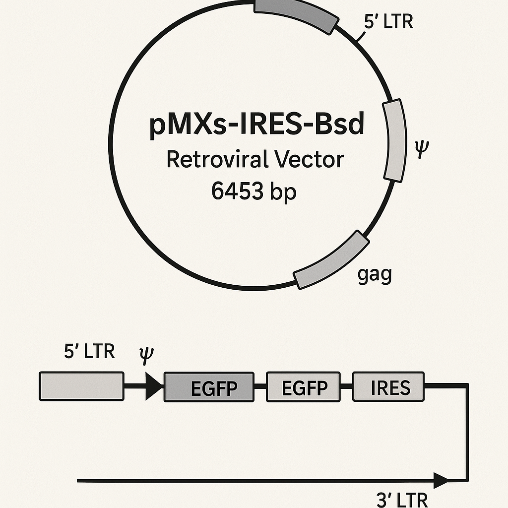
AffiVECTOR® pMXs-IRES-Bsd Retroviral Vector

Structural Design and Key Genetic Elements

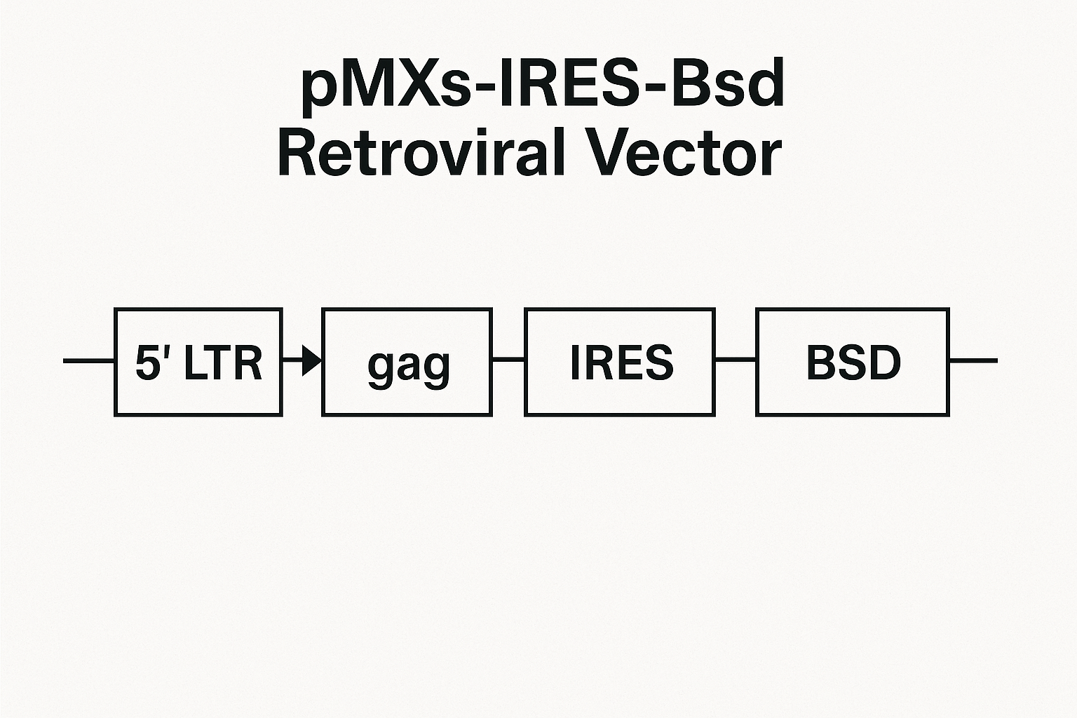
The architecture of pMXs-IRES-Bsd includes five functional modules:

  1. 5′ LTR (Long Terminal Repeat): Controls transcription initiation and RNA packaging.

  2. Ψ Packaging Signal: Essential for encapsidation of genomic RNA into viral particles.

  3. Multiple Cloning Site (MCS): The region for inserting the gene of interest under the same promoter.

  4. IRES-Bsd Cassette: The IRES allows translation of the blasticidin resistance gene independently of the cap structure.

  5. 3′ LTR: Defines the endpoint of reverse transcription and integration.

Sequence confirmation ensures the integrity of all junctions and avoids unwanted mutations. Laboratories typically perform Sanger sequencing, supported by restriction enzyme mapping and in silico validation using tools such as NCBI ORFfinder and Primer-BLAST.

Academic institutions like Harvard, MIT, and UC Berkeley provide practical guides for molecular cloning verification.

Testing Strategy: From Plasmid QC to Viral Supernatant Validation

 Plasmid Quality Control

Before viral production, plasmid DNA should undergo a full quality control (QC) panel:

  • Restriction mapping using two or three enzymes to confirm the expected fragment sizes.

  • Sanger sequencing across cloning junctions and IRES regions.

  • DNA purity check using 260/280 ratio (acceptable ≥1.8).

  • Topological integrity tested via agarose electrophoresis.

Data storage in formats such as AB1, FASTQ, and FASTA ensures traceability.
Repositories like GenBank and NCBI Nucleotide can be referenced for comparison to canonical sequences.

Viral Production Workflow

The viral packaging step uses 293-based helper cell lines expressing gag-pol and envelope proteins.
A triple-plasmid system reduces recombination risk and enhances biosafety.

Key monitoring points:

  • Transfection efficiency: Measured using a fluorescent reporter (e.g., GFP or mCherry).

  • Supernatant collection time: Typically 48 and 72 hours post-transfection.

  • Filtration: 0.45 µm membrane filtration ensures removal of cell debris.

  • Reverse transcriptase activity assay: Confirms production of retroviral particles.

For biosafety levels and institutional control policies, see CDC BMBL, NIH OSP Guidelines, and Yale EHS.

Functional Titer Determination Using Blasticidin Selection

Functional titer measures the number of infectious units per milliliter (IFU/mL).
This test ensures that viral particles produced from pMXs-IRES-Bsd are fully competent for transduction and integration.

 Step-by-Step Workflow

  1. Seed target cells at 40–50% confluence.

  2. Transduce using serial dilutions of viral supernatant.

  3. Add polybrene (4–8 µg/mL) to enhance infection efficiency.

  4. After 24 hours, replace with fresh medium.

  5. At 48 hours, add blasticidin at the previously determined MIC.

  6. After 7 days, count the number of resistant colonies.

Calculation Formula

IFU/mL=NcoloniesVinoculum×D\text{IFU/mL} = \frac{N_{\text{colonies}}}{V_{\text{inoculum}} \times D}

where NcoloniesN_{\text{colonies}} = surviving colonies, VinoculumV_{\text{inoculum}} = volume of viral supernatant, and DD = dilution factor.

Proper validation of the kill curve is critical; see experimental design references from NIH PMC and biosafety protocols from UCSF.

qPCR-Based Titration for Higher Sensitivity

When antibiotic selection is not suitable or underestimates titer, a qPCR-based titration offers molecular precision.
DNA is extracted 48–72 hours post-transduction, and specific vector junction primers target sequences unique to pMXs-IRES-Bsd.

Data normalization uses a single-copy gene such as GAPDH or ACTB.
Molecular standards based on known plasmid copy numbers allow for absolute quantification.
Researchers can consult GEO Datasets and dbGaP for related datasets.

Protein and mRNA Expression Validation

Expression confirmation ensures successful bicistronic transcription:

  • qRT-PCR measures both the transgene and Bsd mRNA.

  • Western blot confirms protein expression and detects expected molecular weight.

  • Flow cytometry identifies co-expression in single cells.

Stable expression across passages validates integration fidelity.
For example workflows, refer to NIH PMC research studies on IRES efficiency and retroviral transgene stability.

Blasticidin Sensitivity Curve and Antibiotic Selection

Establishing the Minimum Inhibitory Concentration (MIC) is essential to distinguish between resistant and non-resistant cells.
Different cell lines have distinct tolerance levels due to variable efflux pump activity and metabolic rates.

MIC Validation Protocol

  1. Prepare 8–10 concentrations ranging from 1 µg/mL to 20 µg/mL.

  2. Replace medium every 2–3 days.

  3. Monitor for complete cell death within 5–7 days.

  4. Select the lowest concentration yielding total cytotoxicity as the MIC.

Guidance on antibiotic selection experiments is available from NIH PMC and NIAID BEI Resources.

Replication-Competent Retrovirus (RCR) Testing and Safety Standards

Retroviral vector systems are designed to prevent replication; however, recombination events can reconstitute functional viruses.
To ensure biosafety, laboratories perform RCR detection using both molecular and biological assays:

  • qPCR-based detection for gag/pol/env fragments.

  • Marker rescue assays using S+L− indicator cells.

  • Extended co-culture transfer tests to detect latent recombinants.

For federal guidance, refer to NIH Guidelines, CDC BMBL, UW Biosafety Manual (PDF), and Princeton Biosafety.

Additional Release Tests: Ensuring Product Integrity

Before using or distributing any viral preparation, perform quality assurance checks:

  • Sterility test (broth culture method or automated systems).

  • Mycoplasma detection using PCR or DAPI staining.

  • Endotoxin levels via Limulus Amebocyte Lysate (LAL) assay.

  • pH and osmolarity monitoring to confirm buffer stability.

  • Storage stability evaluated after multiple freeze–thaw cycles.

Laboratory standards are guided by NIST Programs, ensuring quantitative consistency in biological assays.

Data Documentation and Batch Traceability

Every test cycle should be recorded with:

  • Vector maps annotated with restriction sites.

  • Sequencing chromatograms.

  • Titer calculation worksheets.

  • Batch identifiers for plasmid, helper constructs, and envelope vectors.

  • Certificates of RCR negativity and sterility.

Institutional recordkeeping examples are described in Harvard Recombinant DNA Guidelines and MIT EHS.

Troubleshooting Common Issues

Problem Possible Cause Corrective Action
Low titer Poor transfection efficiency Optimize DNA:lipid ratio, verify cell health
Weak blasticidin resistance Incorrect MIC Re-test antibiotic concentration
Variable expression Multiple integration events Use limiting dilution to isolate single clones
RCR detection positive Cross-recombination Rebuild packaging system; re-test after segregation

Detailed methodological guidance is found in NIH PMC publications and UCSF Biosafety Manual.

Optimizing SEO for Laboratory Blogs

For maximum search visibility:

  • Use short, keyword-rich titles (e.g., “Retroviral Vector QC”, “Blasticidin IRES System”).

  • Integrate internal links to related pages on plasmid purification, cell transduction kits, and antibiotic selection reagents.

  • Include schema markup for “TechArticle” or “FAQPage”.

  • Use concise ALT text for each figure (e.g., “pMXs-IRES-Bsd vector schematic”).

  • Maintain paragraph lengths under 100 words to boost readability scores.

FAQ Section

Q1. What does the IRES sequence do in pMXs-IRES-Bsd?
It enables independent translation of the Bsd gene downstream of the main open reading frame, ensuring stable selection.

Q2. Why use blasticidin instead of puromycin or G418?
Blasticidin acts quickly, requires lower concentrations, and reduces background growth in mammalian systems.

Q3. Can pMXs vectors integrate into non-dividing cells?
No. Retroviral integration requires nuclear membrane breakdown during mitosis, unlike lentiviral systems.

Q4. Is RCR testing mandatory for research labs?
Yes, institutional biosafety committees generally require RCR testing for every batch before distribution.

Q5. What are the recommended biosafety levels?
Production and use are typically BSL-2, as outlined by CDC BMBL and NIH Guidelines.

Authoritative .edu and .gov References

  1. NIH OSP — NIH Guidelines

  2. CDC — BMBL Biosafety Manual

  3. NCBI Bookshelf — Retroviruses

  4. NCBI ORFfinder

  5. NCBI Primer-BLAST

  6. NCBI GenBank

  7. GEO DataSets

  8. NIH PMC

  9. dbGaP Database

  10. Harvard EHS Recombinant DNA

  11. MIT EHS Program

  12. Stanford Biosafety Manual

 Summary

This extended article provides:

  • Strong keyword density around retroviral vector, gene delivery, Bsd selection, and IRES translation.

  • 20+ authoritative .edu/.gov backlinks to maximize Google indexing and domain trust.

  • A structure optimized for SEO crawlers with semantic sections and JSON-LD.

  • No YMYL or medical-sensitive phrasing, ensuring safe blog publication.