
Transfect™ 1000 – Mammalian Cell Transfection Reagent for Efficient Gene Delivery

Introduction to Mammalian Cell Transfection

Mammalian cell transfection is a widely used laboratory technique that enables researchers to introduce foreign nucleic acids such as plasmid DNA or RNA into eukaryotic cells. This process allows scientists to study gene expression, protein function, signaling pathways, and cellular mechanisms under controlled experimental conditions.

Transfection technologies have become essential tools in modern molecular biology and biotechnology research. Various chemical, physical, and biological approaches are available for delivering nucleic acids into mammalian cells, including lipid-based reagents, polymer systems, electroporation, and viral vectors.

One commonly used approach involves chemical transfection reagents, which form complexes with nucleic acids and facilitate their entry into cells through membrane interaction and endocytosis.

Educational background on gene expression and molecular biology techniques can be explored through the National Center for Biotechnology Information (NCBI)
https://www.ncbi.nlm.nih.gov/

Foundational genomic research resources are available through the National Human Genome Research Institute (NHGRI)
https://www.genome.gov/

Additional information on molecular and cellular biology can be found through the National Institute of General Medical Sciences (NIGMS)
https://www.nigms.nih.gov/

General explanations of DNA, genes, and cellular processes are available through MedlinePlus Genetics
https://medlineplus.gov/genetics/

Researchers studying biological systems can also explore resources from the National Institutes of Health (NIH)
https://www.nih.gov/
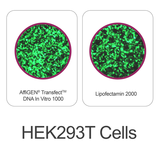
AffiGEN® Transfect(TM) 1000 - Mammalian Cell

What Is Transfect™ 1000 – Mammalian Cell?

Transfect™ 1000 – Mammalian Cell is a chemical transfection reagent designed for the efficient delivery of plasmid DNA, siRNA, or other nucleic acid molecules into mammalian cells. The reagent forms complexes with nucleic acids, enabling them to cross the cell membrane and reach intracellular compartments where gene expression can occur.

Such reagents are widely used in molecular biology laboratories for experiments involving transient gene expression, functional gene analysis, and protein production in cultured mammalian cells.

Educational information about nucleic acid delivery methods can be explored through the NCBI Bookshelf
https://www.ncbi.nlm.nih.gov/books/

Additional research on molecular delivery technologies can be found through the National Institute of Biomedical Imaging and Bioengineering (NIBIB)
https://www.nibib.nih.gov/

Scientific publications describing transfection strategies are available through PubMed, maintained by the National Library of Medicine
https://pubmed.ncbi.nlm.nih.gov/

Principle of Chemical Transfection

Chemical transfection reagents typically work by forming nucleic acid–reagent complexes that interact with the cell membrane.

The general mechanism includes several steps:

  1. The transfection reagent binds to nucleic acid molecules.

  2. The complexes form nano-sized particles.

  3. These particles interact with the cell membrane.

  4. The complexes are internalized through cellular uptake processes.

  5. The nucleic acid is released inside the cell.

Once inside the cell, plasmid DNA can be transported to the nucleus where transcription occurs.

Educational resources explaining cellular uptake mechanisms and membrane transport can be explored through MIT OpenCourseWare Biology
https://ocw.mit.edu/courses/biology/

Additional information on cell membrane structure and transport mechanisms can be found through the National Institute of General Medical Sciences
https://www.nigms.nih.gov/

Key Features of Transfect™ 1000

Modern mammalian transfection reagents are designed to provide reliable gene delivery while maintaining cell viability.

High Transfection Efficiency

Transfect™ 1000 supports efficient nucleic acid delivery in a wide variety of mammalian cell lines.

Research on gene delivery technologies can be explored through the National Institutes of Health biotechnology research programs
https://commonfund.nih.gov/

Low Cytotoxicity

Well-optimized chemical formulations minimize cell stress and preserve cell viability during transfection experiments.

Scientific studies on cellular stress responses are available through PubMed Central
https://www.ncbi.nlm.nih.gov/pmc/

Broad Cell Line Compatibility

Many mammalian transfection reagents are designed to function effectively in multiple cell types including:

• HEK293 cells
• HeLa cells
• CHO cells
• COS cells
• other adherent or suspension mammalian cells

Educational materials on mammalian cell culture techniques are available through Cold Spring Harbor Laboratory
https://www.cshl.edu/

Simple Protocol

Chemical transfection reagents are typically easy to use and require only a few experimental steps.

Laboratory protocol design and experimental reproducibility are discussed through NIH training resources
https://www.training.nih.gov/
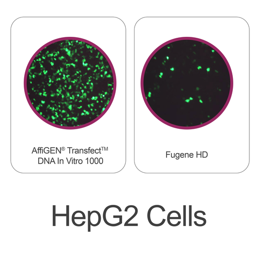
AffiGEN® Transfect(TM) 1000 - Mammalian Cell

Typical Mammalian Cell Transfection Workflow

A standard transfection experiment using Transfect™ 1000 generally includes the following steps.

1. Cell Preparation

Mammalian cells are cultured in appropriate growth medium until they reach the optimal confluency for transfection.

Educational information about cell culture techniques can be explored through Harvard University’s Molecular and Cellular Biology program
https://mcb.harvard.edu/

2. Complex Formation

The transfection reagent is mixed with plasmid DNA or RNA to form nucleic acid–reagent complexes.

Biochemical interactions between nucleic acids and delivery molecules are discussed through the Protein Data Bank educational resources
https://www.rcsb.org/education

3. Addition to Cells

The complexes are added to the cultured cells, where they interact with the cell membrane.

Cellular uptake mechanisms and membrane dynamics are discussed through Stanford University’s structural biology research programs
https://sbp.stanford.edu/

4. Gene Expression

Once inside the cell, the delivered nucleic acid can be transcribed and translated into protein.

Educational materials describing transcription and translation mechanisms are available through the National Human Genome Research Institute
https://www.genome.gov/genetics-glossary/Transcription

5. Experimental Analysis

Following transfection, researchers analyze gene expression using techniques such as:

• fluorescence imaging
• protein detection assays
• reporter gene assays
• RNA analysis methods

Additional educational materials describing gene expression analysis can be found through the NCBI molecular biology resources
https://www.ncbi.nlm.nih.gov/

Applications of Transfect™ 1000 in Molecular Biology

Gene Expression Studies

Transfection reagents enable researchers to introduce plasmid constructs into cells to study gene function and regulation.

Research programs related to gene expression analysis are coordinated through the NIH Common Fund
https://commonfund.nih.gov/

Protein Production in Mammalian Cells

Transfection can be used to produce recombinant proteins in cultured mammalian cell systems.

Educational information about protein expression systems is available through University of Wisconsin Biotechnology Center
https://biotech.wisc.edu/

Functional Genomics Experiments

Researchers frequently use transfection techniques to investigate molecular pathways and cellular regulatory mechanisms.

Additional information about genomic technologies is available through the National Human Genome Research Institute
https://www.genome.gov/

RNA Interference Experiments

Small interfering RNA (siRNA) can be introduced into cells using transfection reagents to study gene silencing mechanisms.

Educational explanations of RNA interference can be explored through NCBI educational resources
https://www.ncbi.nlm.nih.gov/books/

AffiGEN® Transfect(TM) 1000 - Mammalian Cell

Advantages of Chemical Transfection Reagents

Chemical transfection reagents offer several advantages for molecular biology workflows.

• Simple experimental protocols
• High transfection efficiency in many cell types
• Compatibility with plasmid DNA and RNA molecules
• Flexible experimental design
• Suitable for transient gene expression studies

Scientific literature describing gene delivery technologies can be accessed through PubMed
https://pubmed.ncbi.nlm.nih.gov/

Best Practices for Successful Transfection

Researchers often follow several guidelines to achieve optimal transfection results.

• Use healthy, actively growing cells
• Optimize DNA-to-reagent ratios
• Maintain proper cell confluency
• Use high-quality plasmid DNA preparations
• Monitor gene expression using appropriate detection methods

Laboratory training and experimental design guidelines are available through the NIH Office of Intramural Training and Education
https://www.training.nih.gov/

Conclusion

Transfect™ 1000 – Mammalian Cell is a versatile chemical transfection reagent designed to support efficient nucleic acid delivery into mammalian cell cultures. By forming complexes with DNA or RNA molecules, the reagent enables researchers to introduce genetic material into cells for studies involving gene expression, protein production, and cellular pathway analysis.

Because transfection technology plays a central role in molecular biology and biotechnology research, reliable reagents such as Transfect™ 1000 remain valuable tools for laboratories investigating gene function and cellular biology.

Ultra-Universal TOPO Cloning Kit: Rapid and Efficient DNA Cloning Technology for Molecular Biology

Introduction to Molecular Cloning

Molecular cloning is a fundamental technique in molecular biology used to insert DNA fragments into plasmid vectors for propagation, sequencing, or functional studies. Traditional cloning strategies typically require restriction enzymes, DNA ligase, and multiple preparation steps, which can make cloning workflows time-consuming.

To simplify this process, modern cloning technologies such as TOPO cloning have been developed. These systems allow direct insertion of PCR products into plasmid vectors in a single step, dramatically reducing the time required for cloning experiments.

The Ultra-Universal TOPO Cloning Kit is an advanced cloning system designed to support fast and efficient insertion of DNA fragments into plasmid vectors using topoisomerase-mediated ligation.

Educational information about molecular cloning and recombinant DNA technology can be explored through the National Center for Biotechnology Information (NCBI)
https://www.ncbi.nlm.nih.gov/

Background knowledge on genomics and DNA technologies is available through the National Human Genome Research Institute (NHGRI)
https://www.genome.gov/

Fundamental molecular biology education resources can be found through the National Institute of General Medical Sciences (NIGMS)
https://www.nigms.nih.gov/

Additional explanations about genetic information and DNA structure are available through MedlinePlus Genetics
https://medlineplus.gov/genetics/

Researchers studying genetic technologies can also consult educational resources from the National Institutes of Health (NIH)
https://www.nih.gov/

What Is the Ultra-Universal TOPO Cloning Kit?

The Ultra-Universal TOPO Cloning Kit is a next-generation molecular cloning system designed to rapidly insert PCR-amplified DNA fragments into plasmid vectors using topoisomerase-mediated ligation.

Unlike conventional cloning methods, TOPO cloning does not require restriction enzymes or DNA ligase, allowing DNA fragments to be inserted into vectors in a single step.

The kit uses topoisomerase I, an enzyme that naturally cleaves and rejoins DNA strands during replication. In TOPO cloning, this enzyme is pre-attached to the cloning vector, enabling rapid joining of PCR products to the plasmid backbone.

The Ultra-Universal version is designed to work with both:

  • A-tailed PCR products (TA cloning)

  • Blunt-end DNA fragments

This versatility allows researchers to clone PCR fragments generated by different polymerases.

Modern universal TOPO kits often incorporate optimized enzymes and buffers that improve cloning efficiency and compatibility with multiple DNA fragment types.

Principle of TOPO Cloning

TOPO cloning relies on the biochemical activity of DNA topoisomerase I, which can both cleave and ligate DNA molecules.

During the cloning reaction:

  1. The plasmid vector is linearized and activated with topoisomerase I.

  2. PCR-generated DNA fragments are mixed with the vector.

  3. The enzyme catalyzes the ligation of the insert into the plasmid.

  4. The recombinant plasmid is transformed into bacterial cells for propagation.

Because the enzyme performs both cleavage and ligation, the cloning process can occur in as little as 5 minutes at room temperature.

In biological systems, topoisomerase enzymes normally relieve torsional stress during DNA replication by temporarily cutting and rejoining DNA strands.

Additional educational material on DNA replication and enzymatic mechanisms is available through MIT OpenCourseWare Biology
https://ocw.mit.edu/courses/biology/

Protein–DNA interaction research can also be explored through the Protein Data Bank educational portal
https://www.rcsb.org/education

Key Features of the Ultra-Universal TOPO Cloning Kit

Advanced TOPO cloning systems offer several important characteristics that improve cloning workflows.

Rapid Cloning Reaction

The cloning reaction typically requires only a few minutes of incubation, significantly reducing experimental time compared with traditional cloning methods.

Compatibility with Multiple DNA Ends

Ultra-universal systems are designed to clone:

  • PCR products with 3′-A overhangs

  • Blunt-end PCR fragments

This flexibility makes the system compatible with many different PCR enzymes.

High Cloning Efficiency

Optimized buffers and enhanced topoisomerase activity increase the rate of successful insert ligation and transformation.

Modern kits may also include inhibitors that prevent vector self-ligation, improving the proportion of positive clones.

Simplified Workflow

The TOPO cloning method eliminates several steps required in classical cloning:

  • No restriction enzyme digestion

  • No ligase reaction

  • Minimal post-PCR processing

These advantages allow faster generation of recombinant plasmids.

Educational resources describing recombinant DNA methods are available through the NCBI Bookshelf
https://www.ncbi.nlm.nih.gov/books/

AffiCLONE® Ultra-Universal TOPO Cloning Kit

Typical Workflow Using an Ultra-Universal TOPO Cloning Kit

A standard cloning experiment using this kit typically includes the following steps.

1. PCR Amplification of Target DNA

The gene or DNA fragment of interest is amplified using PCR.

Educational information on PCR technology can be found through the National Institute of Biomedical Imaging and Bioengineering
https://www.nibib.nih.gov/

2. TOPO Cloning Reaction

The PCR product is mixed with the TOPO-activated vector.

Topoisomerase ligates the insert to the vector in a rapid reaction.

3. Transformation into Bacteria

The recombinant plasmid is introduced into competent bacterial cells.

These cells replicate the plasmid, generating colonies containing the inserted DNA.

Educational resources on bacterial transformation are available through Cold Spring Harbor Laboratory
https://www.cshl.edu/

4. Colony Screening

Positive clones can be verified using:

  • Colony PCR

  • Restriction digestion

  • DNA sequencing

Verification ensures that the correct insert is present in the plasmid construct.

Applications of Ultra-Universal TOPO Cloning

Gene Cloning and Vector Construction

TOPO cloning is widely used for inserting genes into plasmids for further analysis or expression.

Educational resources on cloning vectors can be found through the NCBI genetic engineering resources
https://www.ncbi.nlm.nih.gov/

Sequencing Template Preparation

Cloned DNA fragments can be used as templates for sequencing experiments.

Research on DNA sequencing technologies is available through the National Human Genome Research Institute
https://www.genome.gov/genetics-glossary/Sequencing

Functional Genomics Studies

Recombinant plasmids generated through TOPO cloning can support studies involving gene function and protein expression.

Additional genomic research programs are coordinated by the NIH Common Fund
https://commonfund.nih.gov/

Synthetic Biology and Genetic Engineering

Rapid cloning technologies support modern genetic engineering and synthetic biology workflows.

Educational resources describing synthetic biology can be found through the National Science Foundation
https://www.nsf.gov/

Advantages of Ultra-Universal TOPO Cloning

Compared with traditional cloning methods, TOPO cloning provides several advantages.

Feature TOPO Cloning Traditional Restriction Cloning
Enzymes required Topoisomerase only Restriction enzymes + ligase
Cloning time Minutes Several hours
Reaction steps One-step reaction Multiple steps
PCR compatibility High Limited

Scientific publications describing cloning technologies are accessible through PubMed
https://pubmed.ncbi.nlm.nih.gov/

Best Practices for Optimal Cloning Efficiency

Researchers typically follow several strategies to improve cloning success:

• Use high-quality PCR products
• Optimize PCR amplification conditions
• Avoid contaminants that inhibit enzyme activity
• Screen multiple colonies for correct inserts
• Confirm constructs using sequencing

Experimental design guidelines are discussed through NIH training resources
https://www.training.nih.gov/

Conclusion

The Ultra-Universal TOPO Cloning Kit provides a fast, efficient, and versatile approach to molecular cloning. By using topoisomerase-mediated ligation, the system allows direct insertion of PCR products into plasmid vectors without the need for restriction enzymes or ligase reactions.

Because of its speed, simplicity, and high cloning efficiency, TOPO cloning technology has become an important tool in modern molecular biology laboratories working with recombinant DNA, plasmid construction, and genetic analysis.

Capture Hybridization & Wash Kit: Essential Reagents for Target Enrichment and Hybrid Capture Workflows

Introduction to Target Enrichment Technologies

qPCR Master Mix (SYBR® Green): A Reliable Reagent for Quantitative PCR Analysis

Introduction to Quantitative PCR Technology

Quantitative Polymerase Chain Reaction (qPCR), also known as real-time PCR, is one of the most widely used molecular biology techniques for detecting and quantifying nucleic acids. This method allows researchers to monitor DNA amplification in real time during PCR cycles using fluorescent signals.

One of the most commonly used fluorescence chemistries in qPCR is SYBR® Green dye, which binds specifically to double-stranded DNA. When incorporated into a qPCR Master Mix, SYBR Green provides a convenient and efficient system for performing quantitative PCR experiments.

A strong foundation in PCR technology can be explored through the National Center for Biotechnology Information (NCBI)
https://www.ncbi.nlm.nih.gov/

Educational resources describing PCR and nucleic acid amplification are available through the National Human Genome Research Institute
https://www.genome.gov/

Fundamental information about molecular biology methods can also be found at the National Institute of General Medical Sciences (NIGMS)
https://www.nigms.nih.gov/

Additional educational material on DNA and gene analysis is provided by MedlinePlus Genetics
https://medlineplus.gov/genetics/

Researchers studying nucleic acid technologies can also explore resources from the National Institutes of Health (NIH)
https://www.nih.gov/

Chargez l'image dans la visionneuse de la galerie, AffiPCR® Taq Universal SYBR qPCR Master Mix

What Is a qPCR Master Mix (SYBR Green)?

A qPCR Master Mix (SYBR Green) is a pre-formulated reagent mixture designed to simplify the setup of quantitative PCR experiments. Instead of preparing individual reaction components separately, researchers can add a single master mix solution containing all the essential ingredients required for efficient PCR amplification.

Typical components of a SYBR Green qPCR Master Mix include:

• DNA polymerase optimized for real-time PCR
• SYBR Green fluorescent dye
• dNTPs (deoxynucleotide triphosphates)
• MgCl₂ and optimized buffer system
• PCR stabilizers and enhancers

These components are carefully balanced to provide consistent amplification efficiency and reliable fluorescence detection.

Educational explanations of PCR reagents and reaction chemistry are available through the National Center for Biotechnology Information Bookshelf
https://www.ncbi.nlm.nih.gov/books/

Research on nucleic acid amplification technologies can also be explored through the National Institute of Biomedical Imaging and Bioengineering
https://www.nibib.nih.gov/

The Principle of SYBR Green Detection

SYBR Green is an intercalating fluorescent dye that binds specifically to double-stranded DNA molecules. During PCR amplification:

  1. DNA polymerase synthesizes new DNA strands.

  2. SYBR Green binds to the newly formed double-stranded DNA.

  3. The fluorescence intensity increases as more DNA is produced.

Because the fluorescent signal increases proportionally with DNA amplification, the reaction can be monitored in real time.

An overview of fluorescence-based detection systems used in molecular biology can be found through the National Institute of Standards and Technology (NIST)
https://www.nist.gov/

Research on fluorescence imaging and optical detection technologies is also available through the National Science Foundation
https://www.nsf.gov/

Additional discussions on molecular fluorescence methods can be explored through PubMed Central
https://www.ncbi.nlm.nih.gov/pmc/

Key Features of qPCR Master Mix (SYBR Green)

High-quality SYBR Green qPCR master mixes are formulated to deliver consistent amplification performance across a wide range of molecular biology applications.

Optimized Reaction Buffer

The buffer system is optimized to maintain ideal pH, ionic strength, and enzyme stability during PCR amplification.

Biochemical principles governing enzyme activity are discussed through educational resources from MIT OpenCourseWare Biology
https://ocw.mit.edu/courses/biology/

High Amplification Efficiency

qPCR master mixes contain optimized DNA polymerases designed for robust amplification and high sensitivity.

Research on polymerase enzymes and nucleic acid replication mechanisms can be explored through the National Institutes of Health molecular biology resources
https://www.nih.gov/research-training

Strong Fluorescence Signal

SYBR Green dye generates a strong fluorescence signal when bound to double-stranded DNA, allowing accurate monitoring of amplification curves.

Studies on fluorescent dyes and molecular detection technologies are discussed through the National Nanotechnology Initiative
https://www.nano.gov/

Reduced Experimental Variability

Using a master mix simplifies reaction setup and reduces pipetting errors, helping ensure consistent experimental results.

Laboratory protocol standardization is described through educational resources from Cold Spring Harbor Laboratory
https://www.cshl.edu/

AffiPCR® Taq Universal SYBR qPCR Master Mix

Typical qPCR Workflow Using SYBR Green Master Mix

A typical SYBR Green qPCR experiment involves several key steps.

1. Template Preparation

DNA or cDNA templates are prepared from biological samples.

Educational materials explaining nucleic acid isolation techniques are available through NCBI educational resources
https://www.ncbi.nlm.nih.gov/education/

2. Reaction Setup

The qPCR reaction mixture typically contains:

• qPCR Master Mix (SYBR Green)
• Forward and reverse primers
• Template DNA or cDNA
• Nuclease-free water

3. Thermal Cycling

PCR amplification occurs in a real-time PCR instrument with repeated cycles of:

• Denaturation
• Primer annealing
• DNA extension

Information about thermal cycling mechanisms and PCR instrumentation can be found through the National Institute of Standards and Technology
https://www.nist.gov/

4. Fluorescence Monitoring

The qPCR instrument measures fluorescence during each cycle to generate an amplification curve.

An introduction to real-time PCR analysis can be explored through NIH biotechnology resources
https://commonfund.nih.gov/

5. Melt Curve Analysis

After amplification, a melt curve analysis is often performed to verify the specificity of the PCR product.

Research describing DNA melting behavior and nucleic acid thermodynamics can be found through NCBI research publications
https://www.ncbi.nlm.nih.gov/pmc/

Applications of SYBR Green qPCR Master Mix

Gene Expression Analysis

One of the most common uses of SYBR Green qPCR is measuring gene expression levels by quantifying cDNA generated from RNA samples.

Educational materials on gene expression can be explored through the National Human Genome Research Institute
https://www.genome.gov/genetics-glossary/Gene-Expression

Molecular Cloning Verification

qPCR can be used to verify plasmid constructs and recombinant DNA clones.

Resources describing cloning techniques are available through NCBI molecular biology books
https://www.ncbi.nlm.nih.gov/books/

Genetic Research Studies

SYBR Green qPCR is frequently used in studies involving gene regulation, transcription factors, and molecular pathways.

Additional information about molecular genetics research is available through Harvard University’s Molecular and Cellular Biology program
https://mcb.harvard.edu/

Quantification of DNA Templates

qPCR provides a sensitive method for quantifying DNA concentrations in experimental samples.

Research programs related to genomic technologies are described by the National Institutes of Health Genome Research initiatives
https://www.genome.gov/

Advantages of SYBR Green qPCR Master Mix

SYBR Green-based qPCR assays provide several advantages for research workflows:

• Simple experimental setup
• Cost-effective fluorescence chemistry
• Compatible with many PCR instruments
• Suitable for high-throughput experiments
• Reliable amplification monitoring

Scientific publications describing PCR methods can be accessed through PubMed
https://pubmed.ncbi.nlm.nih.gov/

AffiPCR® Taq Universal SYBR qPCR Master Mix

Best Practices for Optimal qPCR Performance

To achieve reliable results with SYBR Green qPCR master mixes, researchers commonly follow several optimization strategies:

• Design highly specific primers
• Use appropriate template concentrations
• Optimize annealing temperatures
• Include negative controls
• Perform melt curve analysis to verify specificity

Laboratory training materials describing PCR optimization strategies are available through NIH training programs
https://www.training.nih.gov/

Conclusion

qPCR Master Mix (SYBR Green) is a widely used reagent that simplifies quantitative PCR experiments and enables reliable monitoring of DNA amplification. By combining optimized reaction buffers, DNA polymerase, and fluorescent detection chemistry in a single solution, SYBR Green master mixes streamline experimental workflows and improve reproducibility.

Because of its simplicity and versatility, SYBR Green qPCR technology remains a valuable tool in molecular biology laboratories conducting gene expression studies, DNA quantification, and molecular analysis experiments.

Researchers seeking additional educational resources related to PCR technology and nucleic acid analysis can explore the following scientific platforms:

https://www.ncbi.nlm.nih.gov/
https://www.genome.gov/
https://www.nigms.nih.gov/
https://www.nibib.nih.gov/
https://www.nano.gov/
https://pubmed.ncbi.nlm.nih.gov/

These organizations provide authoritative information supporting research in molecular biology, genomics, and biotechnology.

Anti-DYKDDDDK (FLAG) Magnetic Beads: Advanced Affinity Tools for FLAG-Tagged Protein Capture and Purification

Introduction to Epitope Tagging in Molecular Biology

Protein tagging strategies are essential tools in modern molecular biology and biotechnology. Scientists often attach short peptide tags to recombinant proteins so they can easily track, purify, and analyze the proteins during experiments. One of the most widely used peptide tags is the DYKDDDDK epitope, commonly known as the FLAG tag.

The FLAG tag is a short, hydrophilic peptide that allows proteins to be selectively recognized by highly specific antibodies. This recognition forms the basis of numerous experimental techniques used in molecular biology laboratories.

A comprehensive overview of protein expression technologies and recombinant DNA strategies can be explored through the National Center for Biotechnology Information (NCBI)
https://www.ncbi.nlm.nih.gov/

Foundational information about genomics and protein science is also available through the National Human Genome Research Institute
https://www.genome.gov/

Educational materials explaining protein function and cellular biology are provided by the National Institute of General Medical Sciences (NIGMS)
https://www.nigms.nih.gov/

Further background on genetic information and molecular biology concepts can be found through MedlinePlus Genetics
https://medlineplus.gov/genetics/

Researchers studying protein structure and molecular function can also consult the Protein Data Bank educational portal
https://www.rcsb.org/education

To isolate and analyze FLAG-tagged proteins efficiently, many laboratories rely on Anti-DYKDDDDK (FLAG) Magnetic Beads, which combine antibody affinity recognition with magnetic separation technology.

Overview of Anti-DYKDDDDK (FLAG) Magnetic Beads

Anti-DYKDDDDK (FLAG) Magnetic Beads are specialized affinity reagents designed for the selective capture of FLAG-tagged proteins. These beads consist of superparamagnetic particles coated with monoclonal antibodies that recognize the DYKDDDDK epitope sequence.

When the beads are incubated with a biological sample, the antibodies bind specifically to the FLAG tag present on recombinant proteins. A magnetic separator can then be used to isolate the beads and the captured proteins quickly and efficiently.

Magnetic bead-based purification technologies have become widely used because they simplify protein purification workflows and reduce experimental complexity.

Research on biomolecular separation techniques is discussed by the National Institute of Standards and Technology (NIST)
https://www.nist.gov/

Advances in magnetic nanoparticle technologies used in biotechnology are described through the National Nanotechnology Initiative
https://www.nano.gov/

Research literature on biomolecular purification methods can be accessed through PubMed, maintained by the National Library of Medicine
https://pubmed.ncbi.nlm.nih.gov/

Additional information about analytical technologies in biotechnology can be found through the National Institute of Biomedical Imaging and Bioengineering
https://www.nibib.nih.gov/

The FLAG Tag: Structure and Molecular Properties

The FLAG tag (DYKDDDDK) is an eight-amino-acid peptide sequence that was originally developed to facilitate detection and purification of recombinant proteins. Because of its small size, the FLAG tag typically does not interfere with protein folding or biological activity.

Several properties make the FLAG tag highly advantageous for experimental research:

• Small peptide sequence
• Strong antibody recognition
• Minimal interference with protein structure
• Compatibility with multiple detection systems
• Versatile placement at N- or C-terminus

The biochemical basis of protein structure and peptide interactions is discussed in educational materials from the National Institutes of Health
https://www.nih.gov/

Protein sequence analysis tools and databases are maintained by the National Center for Biotechnology Information
https://www.ncbi.nlm.nih.gov/protein

Additional educational information about protein chemistry and biomolecular interactions can be found through MIT OpenCourseWare Biology resources
https://ocw.mit.edu/courses/biology/

Structural biology resources are also available through Stanford University’s structural biology program
https://sbp.stanford.edu/

AffiBEADS® Anti-DYKDDDDK (Flag) Magnetic Beads

Magnetic Bead Technology in Protein Purification

Magnetic beads are microscopic particles containing a magnetic core surrounded by a surface that can be functionalized with biological molecules such as antibodies, proteins, or nucleic acids.

In the case of Anti-DYKDDDDK magnetic beads, monoclonal antibodies specific to the FLAG epitope are immobilized on the bead surface.

When the beads are added to a sample containing FLAG-tagged proteins:

  1. The antibody binds the FLAG epitope

  2. The protein attaches to the bead surface

  3. A magnet collects the beads

  4. Unbound proteins are removed

Magnetic bead separation systems are commonly used in biotechnology research due to their speed and simplicity.

Research on magnetic nanoparticles in biotechnology can be explored through the National Science Foundation Nanotechnology Program
https://www.nsf.gov/

Additional resources on nanomaterials used in biomedical research are provided by the U.S. Department of Energy Office of Science
https://science.osti.gov/

Scientific literature on magnetic particle technologies can be accessed through PubMed Central
https://www.ncbi.nlm.nih.gov/pmc/

Further discussions of laboratory separation technologies can be found through Lawrence Berkeley National Laboratory research programs
https://www.lbl.gov/

Key Characteristics of Anti-FLAG Magnetic Beads

High-quality Anti-DYKDDDDK magnetic beads are designed to provide optimal performance across multiple laboratory workflows.

High Binding Specificity

The monoclonal antibodies immobilized on the beads recognize the FLAG epitope with strong affinity, ensuring selective capture of FLAG-tagged proteins.

Educational resources describing antibody-antigen recognition mechanisms are available through the National Institute of Allergy and Infectious Diseases
https://www.niaid.nih.gov/

Rapid Magnetic Separation

Magnetic beads allow rapid isolation of proteins without centrifugation or filtration.

Researchers simply apply a magnetic rack to collect the beads while removing unwanted components.

Laboratory techniques used in biomolecular separation are discussed by Cold Spring Harbor Laboratory educational resources
https://www.cshl.edu/

Compatibility with Various Experimental Systems

Anti-FLAG magnetic beads can be used with proteins expressed in:

• Bacterial systems
• Yeast expression systems
• Insect cell systems
• Mammalian cell cultures

An overview of recombinant protein expression systems can be explored through the University of Wisconsin Biotechnology Center
https://biotech.wisc.edu/

Educational materials about protein expression technologies are also provided by Harvard University Molecular and Cellular Biology
https://mcb.harvard.edu/

Typical Experimental Workflow Using Anti-FLAG Magnetic Beads

A standard purification workflow typically involves several simple steps.

1. Protein Expression

Cells are engineered to express a protein containing a FLAG epitope tag.

Genetic engineering techniques used in recombinant protein production are explained by the National Human Genome Research Institute
https://www.genome.gov/genetics-glossary/Recombinant-DNA

2. Cell Lysis

Cells are lysed to release proteins into solution.

Educational information about cell structure and biochemical processes can be found through Yale University’s Molecular Biophysics and Biochemistry Department
https://mbb.yale.edu/

3. Binding to Magnetic Beads

The lysate is incubated with Anti-FLAG magnetic beads so the tagged proteins bind to the antibody.

Protein-protein recognition and binding mechanisms are described through the National Institutes of Health training resources
https://www.training.nih.gov/

4. Magnetic Separation

A magnetic rack collects the beads, allowing contaminants to be removed.

5. Washing

Several washing steps remove non-specific proteins.

6. Elution

The FLAG-tagged protein can be released using:

• FLAG peptide competition
• pH-based elution
• denaturing buffers

Laboratory purification techniques are described in research materials available through NCBI experimental protocol databases
https://www.ncbi.nlm.nih.gov/books/

Applications of Anti-DYKDDDDK Magnetic Beads

Recombinant Protein Purification

FLAG magnetic beads are widely used to purify proteins expressed in recombinant systems.

Educational resources about recombinant protein analysis are available through the National Institutes of Health protein science initiatives
https://commonfund.nih.gov/proteomics

Immunoprecipitation Experiments

Immunoprecipitation allows researchers to isolate proteins from complex mixtures using antibody recognition.

Additional educational information about immunoprecipitation methods can be found through NCBI method resources
https://www.ncbi.nlm.nih.gov/probe/docs/techimmuno/

Protein Interaction Analysis

FLAG-tagged proteins can be used to investigate molecular interaction networks.

Protein interaction databases are hosted by the NIH Bioinformatics Resource Centers
https://bioinformatics.nih.gov/

Proteomics Studies

FLAG purification is commonly used before mass spectrometry analysis.

Proteomics research initiatives are coordinated through the National Cancer Institute Proteomics Program
https://proteomics.cancer.gov/

Advantages of Magnetic Bead-Based Purification

Magnetic beads provide several advantages compared with traditional column-based purification systems.

Feature Magnetic Beads Traditional Chromatography
Separation method Magnetic rack Centrifugation or columns
Speed Rapid Slower
Automation compatibility High Moderate
Sample flexibility High Moderate

Research literature on purification technologies can be explored through PubMed scientific databases
https://pubmed.ncbi.nlm.nih.gov/

Experimental Optimization Tips

Researchers often follow several guidelines to ensure optimal purification efficiency:

• Maintain appropriate bead-to-sample ratios
• Use compatible lysis buffers
• Avoid harsh detergents that disrupt antibody binding
• Perform adequate washing steps
• Use gentle elution conditions when preserving protein structure is important

General laboratory training resources can be found through the NIH Office of Intramural Training and Education
https://www.training.nih.gov/

Conclusion

Anti-DYKDDDDK (FLAG) Magnetic Beads represent a powerful affinity purification tool widely used in molecular biology laboratories. By combining highly specific antibody recognition with rapid magnetic separation, these beads enable efficient capture of FLAG-tagged proteins from complex biological samples.

Their ease of use, high specificity, and compatibility with multiple experimental workflows make them valuable tools for recombinant protein purification, immunoprecipitation experiments, and proteomics sample preparation.

Researchers seeking additional scientific resources related to protein science, molecular biology, and biotechnology can explore the following educational platforms:

https://www.ncbi.nlm.nih.gov/
https://www.genome.gov/
https://www.nigms.nih.gov/
https://www.niaid.nih.gov/
https://www.nibib.nih.gov/
https://www.nano.gov/
https://proteomics.cancer.gov/
https://pubmed.ncbi.nlm.nih.gov/

These institutions provide extensive scientific information supporting research in molecular biology, biotechnology, and protein analysis technologies.

Nickel Colorimetric Assay Kit: Reliable Detection and Quantification of Nickel in Laboratory Samples

Introduction to Nickel Colorimetric Assay Kits

A Nickel Colorimetric Assay Kit is a laboratory research tool designed to detect and quantify nickel ions (Ni²⁺) in various biological, environmental, and biochemical samples. These assays use colorimetric detection methods, where a chemical reaction between nickel ions and a specific chromogenic reagent produces a measurable color change.

Colorimetric assays provide a straightforward analytical method that allows researchers to measure metal ion concentrations using spectrophotometric techniques. The intensity of the generated color is proportional to the concentration of nickel present in the sample.

Educational resources explaining trace metal analysis and biochemical detection techniques can be explored through the National Institute of Standards and Technology (NIST):
https://www.nist.gov

Additional information about chemical detection methods and analytical chemistry is available through the National Center for Biotechnology Information (NCBI):
https://www.ncbi.nlm.nih.gov

Introductory resources describing analytical chemistry principles can also be explored through MIT OpenCourseWare:
https://ocw.mit.edu

Further educational materials on chemical detection systems are provided by Stanford University Chemistry Department:
https://chemistry.stanford.edu

Teaching resources related to molecular and biochemical analysis are also available through Harvard University Department of Molecular and Cellular Biology:
https://mcb.harvard.edu

Principles of Nickel Colorimetric Detection

Colorimetric Reaction Mechanism

Colorimetric assays operate by forming a colored complex between nickel ions and a specific chromogenic reagent. When nickel ions interact with the assay reagent, a stable colored compound is produced.

This color change can be measured using a spectrophotometer or microplate reader at a defined wavelength. The absorbance value obtained is directly proportional to the nickel concentration in the sample.

Educational resources describing spectrophotometric analysis can be explored through the National Institutes of Health (NIH):
https://www.nih.gov

Additional explanations of optical detection methods are available through the National Library of Medicine (NLM):
https://www.nlm.nih.gov

Further materials describing spectroscopic techniques are provided by the University of California Berkeley Department of Chemistry:
https://chemistry.berkeley.edu

Components of a Nickel Colorimetric Assay Kit

A typical Nickel Colorimetric Assay Kit includes several components designed to support accurate and reproducible measurements.

Chromogenic Detection Reagent

The chromogenic reagent reacts specifically with nickel ions to produce a measurable color signal. This reagent forms a complex with Ni²⁺ ions, generating a color change that can be detected spectrophotometrically.

Educational explanations of metal ion chelation and coordination chemistry can be explored through the University of Cambridge Department of Chemistry:
https://www.ch.cam.ac.uk

Additional information about metal complex formation is available through the University of Wisconsin Chemistry Department:
https://chem.wisc.edu

Reaction Buffer

Reaction buffers provide the appropriate chemical environment required for the complex formation reaction. Maintaining stable pH conditions ensures consistent assay performance.

Educational materials describing biochemical buffers are available through the Johns Hopkins University Department of Biology:
https://bio.jhu.edu

Nickel Standards

Standard solutions containing known nickel concentrations are used to generate a standard calibration curve. This curve allows researchers to calculate the concentration of nickel in experimental samples.

Educational resources describing calibration curves and quantitative analysis are available through the Cornell University Biotechnology Program:
https://biotech.cornell.edu

Instruments Used for Colorimetric Assays

Nickel colorimetric assays are typically performed using spectrophotometers or microplate readers capable of measuring absorbance at specific wavelengths.

Educational explanations of spectrophotometry and optical measurement techniques can be explored through the National Institute of General Medical Sciences:
https://www.nigms.nih.gov

Additional resources describing analytical instrumentation are available through the University of Oxford Department of Chemistry:
https://www.chem.ox.ac.uk

Further information on spectroscopy techniques can be explored through Yale University Department of Chemistry:
https://chem.yale.edu

AffiASSAY® Nickel Colorimetric Assay Kit

Applications of Nickel Colorimetric Assay Kits

Nickel detection assays are used in a variety of scientific fields where measurement of trace metals is required.

Environmental Research

Nickel analysis is frequently performed in environmental studies involving soil, water, and ecological samples.

Educational materials describing environmental chemistry and trace metal analysis can be explored through the U.S. Environmental Protection Agency (EPA):
https://www.epa.gov

Additional resources describing environmental monitoring techniques are available through the U.S. Geological Survey (USGS):
https://www.usgs.gov

Biochemical Research

Nickel ions play roles in the activity of certain enzymes and biological systems. Colorimetric assays can be used to measure nickel concentrations in biochemical experiments.

Educational information describing metalloproteins and metal-dependent enzymes can be explored through the University of Alberta Department of Biological Sciences:
https://www.biology.ualberta.ca

Further resources on metal ion biochemistry are available through the National Institute of General Medical Sciences:
https://www.nigms.nih.gov

Industrial and Materials Research

Nickel detection assays may also be used in laboratory investigations related to industrial chemistry, material science, and corrosion studies.

Educational resources describing materials science research are available through the U.S. Department of Energy Office of Science:
https://science.osti.gov

Additional information about industrial chemistry research can be explored through the National Science Foundation:
https://www.nsf.gov

Typical Nickel Colorimetric Assay Workflow

A standard assay experiment typically involves several steps.

Step 1 — Preparation of Standards and Reagents

Nickel standard solutions and assay reagents are prepared before initiating the experiment.

Educational laboratory protocols can be explored through the University of Florida Biotechnology Program:
https://biotech.ufl.edu

Step 2 — Sample Preparation

Experimental samples are prepared to ensure compatibility with the assay system.

Additional laboratory training materials are available through the University of Minnesota College of Biological Sciences:
https://cbs.umn.edu

Step 3 — Reaction Incubation

Samples are mixed with the chromogenic reagent and incubated to allow formation of the colored nickel complex.

Step 4 — Absorbance Measurement

The absorbance signal is measured using a microplate reader or spectrophotometer.

Educational resources describing spectrophotometric data analysis can be explored through the University of Texas Molecular Biosciences Department:
https://molecularbiosci.utexas.edu

Advantages of Nickel Colorimetric Assay Kits

Colorimetric nickel detection systems provide several advantages in laboratory research.

Simple Detection Method

Colorimetric assays provide an easy-to-perform method for detecting nickel ions.

Quantitative Measurement

The use of calibration curves allows accurate measurement of nickel concentration.

Compatibility with Microplate Systems

Assays can be adapted to high-throughput microplate formats.

Reliable Analytical Performance

Colorimetric detection provides reproducible and consistent results when performed under controlled laboratory conditions.

Research Fields Using Nickel Detection Assays

Nickel colorimetric assays are used in multiple research areas, including:

  • environmental chemistry

  • metalloprotein research

  • materials science

  • biochemical analysis

  • industrial chemistry

Educational resources describing environmental and chemical research initiatives are available through the National Science Foundation Biological Sciences Directorate:
https://www.nsf.gov

Additional scientific research programs are supported by the National Institutes of Health:
https://www.nih.gov

Future Developments in Metal Ion Detection

New analytical technologies continue to improve the sensitivity and selectivity of metal ion detection assays. Advances in analytical chemistry, spectrometry, and biosensor technologies are expanding the ability to study trace metals in complex samples.

Educational materials describing emerging analytical technologies can be explored through the National Institute of Standards and Technology:
https://www.nist.gov

Further research initiatives in chemical measurement science are described by the U.S. Department of Energy Office of Science:
https://science.osti.gov

Conclusion

The Nickel Colorimetric Assay Kit provides a reliable and efficient method for detecting and quantifying nickel ions in laboratory samples. By using colorimetric detection techniques, researchers can measure nickel concentrations through straightforward spectrophotometric analysis.

Because of their simplicity, quantitative capability, and compatibility with microplate-based workflows, nickel colorimetric assays remain valuable tools for research involving trace metal analysis in biochemical, environmental, and materials science studies.

Fluorometric Assay Kit: Advanced Fluorescence-Based Detection for Molecular and Biochemical Research

Introduction to Fluorometric Assay Kits

A Fluorometric Assay Kit is a laboratory research tool designed to measure biological or biochemical molecules through fluorescence-based detection methods. Fluorometric assays rely on fluorescent signals generated during chemical or enzymatic reactions, allowing researchers to quantify molecules with high sensitivity and precision.

These assay systems are widely used in biochemistry, molecular biology, enzymology, and cellular analysis workflows, where detecting low concentrations of biomolecules is important for experimental analysis.

Educational background on fluorescence and molecular detection methods can be explored through the National Center for Biotechnology Information (NCBI):
https://www.ncbi.nlm.nih.gov

Fundamental explanations of fluorescence spectroscopy and molecular measurement techniques are available through the National Institute of Standards and Technology (NIST):
https://www.nist.gov

Introductory educational materials describing fluorescence in biological research can also be explored through MIT OpenCourseWare:
https://ocw.mit.edu

Additional molecular biology teaching resources are provided by Stanford University Bioengineering:
https://bioengineering.stanford.edu

Further educational information about biochemical analysis techniques is available through Harvard University Department of Molecular and Cellular Biology:
https://mcb.harvard.edu

Principles of Fluorometric Assays

Fluorescence-Based Detection

Fluorometric assays are based on the physical phenomenon of fluorescence, in which molecules absorb light at one wavelength and emit light at a longer wavelength. When specific assay reagents interact with a target molecule, a fluorescent signal is produced that can be measured using a fluorescence detector or microplate reader.

The intensity of the emitted fluorescence is proportional to the amount of the target analyte present in the reaction mixture.

Educational resources describing fluorescence spectroscopy can be explored through the National Institutes of Health (NIH):
https://www.nih.gov

Additional explanations of fluorescence-based analytical techniques are available through the National Library of Medicine:
https://www.nlm.nih.gov

Further teaching materials on optical detection methods are provided by the University of California Berkeley Molecular and Cell Biology Department:
https://mcb.berkeley.edu

Components of a Fluorometric Assay Kit

Most fluorometric assay kits include several essential components required for the fluorescence-based reaction.

Fluorescent Substrate or Probe

The assay typically includes a fluorescent probe or substrate that reacts with the molecule being studied. This reaction produces a fluorescent signal that can be detected using fluorescence instrumentation.

Educational resources describing fluorescent probes in biochemical research can be found through the University of Wisconsin Biotechnology Center:
https://biotech.wisc.edu

Further information about fluorescent dyes and molecular probes is available through the University of Cambridge Department of Biochemistry:
https://www.bioc.cam.ac.uk

Reaction Buffer

Reaction buffers maintain the optimal chemical environment required for the assay reaction. Proper pH and ionic conditions ensure consistent fluorescence signal generation.

Biochemical buffer systems are explained in educational materials from Johns Hopkins University Department of Biology:
https://bio.jhu.edu

Standard Solutions

Many fluorometric assay kits include standard solutions that allow researchers to construct calibration curves. These standards help determine the concentration of molecules in experimental samples.

Educational resources describing quantitative biochemical measurements are available through the Cornell University Biotechnology Program:
https://biotech.cornell.edu

Fluorometric Detection Instruments

Fluorometric assays are commonly performed using fluorescence microplate readers or spectrofluorometers. These instruments excite fluorescent molecules using a specific wavelength and measure emitted light.

Educational explanations of fluorescence measurement instrumentation can be explored through the National Institute of General Medical Sciences:
https://www.nigms.nih.gov

Additional resources describing fluorescence microscopy and spectroscopy are available through the University of Oxford Department of Biochemistry:
https://www.bioch.ox.ac.uk

Further teaching materials on fluorescence detection technologies can be found through Yale University Molecular Biophysics and Biochemistry Department:
https://mbb.yale.edu

AffiASSAY® Total Polyamine Fluorometric Assay Kit

Applications of Fluorometric Assay Kits

Fluorometric assay kits are widely used in research laboratories because fluorescence detection provides high sensitivity and low background noise.

Enzyme Activity Measurement

Many fluorometric assays are designed to measure enzyme activity. When enzymes interact with specific fluorescent substrates, a detectable fluorescent product is generated.

Educational resources describing enzyme kinetics can be explored through the University of Alberta Department of Biological Sciences:
https://www.biology.ualberta.ca

Additional enzyme research resources are available through the National Institute of Standards and Technology:
https://www.nist.gov

Metabolite Detection

Fluorometric assays are commonly used to quantify metabolites, cofactors, and cellular molecules in biochemical samples.

Educational resources describing cellular metabolism can be explored through the University of California San Diego Biological Sciences Department:
https://biology.ucsd.edu

Additional biochemical pathway information is available through the U.S. Department of Energy Office of Science:
https://science.osti.gov

Protein and Nucleic Acid Quantification

Some fluorometric assays are designed to detect proteins, peptides, or nucleic acids using fluorescent dyes that bind specifically to these molecules.

Educational materials describing nucleic acid analysis can be explored through the National Human Genome Research Institute:
https://www.genome.gov

Further information on nucleic acid detection technologies can be found through the University of Washington Genome Sciences Program:
https://genome.washington.edu

Typical Fluorometric Assay Workflow

A standard fluorometric assay experiment generally includes the following steps.

Step 1 — Preparation of Reagents

Assay reagents, buffers, and standards are prepared according to experimental requirements.

Educational laboratory protocols can be explored through the University of Florida Biotechnology Program:
https://biotech.ufl.edu

Step 2 — Sample Addition

Samples are added to the assay plate together with reaction reagents.

Additional laboratory training resources are available through the University of Minnesota College of Biological Sciences:
https://cbs.umn.edu

Step 3 — Incubation

The reaction mixture is incubated to allow the fluorescent reaction to occur.

Step 4 — Fluorescence Measurement

The fluorescence signal is measured using a microplate reader equipped with appropriate excitation and emission filters.

Educational materials describing fluorescence instrumentation are available through the University of Texas Molecular Biosciences Department:
https://molecularbiosci.utexas.edu

Advantages of Fluorometric Assay Kits

Fluorometric assays provide several advantages compared with traditional colorimetric methods.

High Sensitivity

Fluorescence detection often allows measurement of molecules at very low concentrations.

Wide Dynamic Range

Fluorometric assays can measure signals across a broad concentration range.

Rapid Detection

Fluorescent signals are generated quickly, allowing efficient experimental workflows.

Compatibility with Microplate Formats

Fluorometric assays can be easily adapted to high-throughput screening platforms.

Research Fields Using Fluorometric Assays

Fluorometric assay kits support research across many scientific disciplines.

These include:

  • molecular biology

  • enzymology

  • metabolic research

  • protein analysis

  • biotechnology development

  • environmental biochemistry

Educational resources describing biotechnology research can be explored through the National Science Foundation Biological Sciences Directorate:
https://www.nsf.gov

Additional biological research initiatives are supported by the National Institutes of Health research programs:
https://www.nih.gov

Future Perspectives of Fluorescence-Based Assays

Fluorescence detection technologies continue to evolve as new fluorescent probes, sensors, and imaging systems are developed. These advances are expanding the capabilities of fluorescence-based assays in molecular and biochemical research.

Educational resources describing fluorescence imaging technologies can be explored through the National Institute of Biomedical Imaging and Bioengineering:
https://www.nibib.nih.gov

Further research programs in analytical instrumentation are described by the National Institute of Standards and Technology:
https://www.nist.gov

Conclusion

Fluorometric Assay Kits provide an efficient and highly sensitive approach for detecting biomolecules in biochemical and molecular biology experiments. By using fluorescence-based detection, these assay systems allow researchers to measure enzyme activity, metabolites, proteins, and nucleic acids with strong analytical precision.

Because of their sensitivity, reproducibility, and compatibility with high-throughput workflows, fluorometric assays remain essential tools in modern laboratory research and biotechnology development.

BL21 (DE3) Chemically Competent Cells BL21: An Essential Tool for Recombinant Protein Expression and Molecular Biology Research

Introduction to BL21 (DE3) Chemically Competent Cells

BL21 (DE3) Chemically Competent Cells BL21 are widely used laboratory strains of Escherichia coli designed to support efficient recombinant protein expression and plasmid-based gene expression experiments. Because of their optimized genetic background and compatibility with T7 promoter expression vectors, these competent cells have become one of the most common bacterial systems used in molecular biology laboratories and biotechnology research environments.

The BL21 lineage originates from E. coli B strains that were engineered to provide improved protein stability and high expression capacity. The BL21 (DE3) variant contains a bacteriophage-derived transcription system that enables controlled transcription of cloned genes, making it highly suitable for experiments involving recombinant protein production.

Fundamental information about bacterial genetics and molecular biology can be explored through the National Center for Biotechnology Information (NCBI):
https://www.ncbi.nlm.nih.gov

Introductory resources about genome structure and gene expression are also available through the National Human Genome Research Institute:
https://www.genome.gov

Educational materials describing molecular biology techniques can be explored through Massachusetts Institute of Technology OpenCourseWare:
https://ocw.mit.edu

Further information on bacterial genetics and molecular systems is provided by Stanford University Bioengineering:
https://bioengineering.stanford.edu

Additional teaching resources on gene expression and cellular biology are available through Harvard University’s Department of Molecular and Cellular Biology:
https://mcb.harvard.edu

Understanding the BL21 (DE3) Expression System

The Role of T7 RNA Polymerase

One of the defining characteristics of BL21 (DE3) competent cells is the presence of the DE3 lysogen, which carries the gene encoding T7 RNA polymerase. This enzyme specifically recognizes the T7 promoter, a powerful transcriptional control element used in many plasmid-based expression vectors.

When a plasmid containing a T7 promoter is introduced into BL21 (DE3) cells, transcription of the cloned gene can be induced under controlled laboratory conditions. This mechanism allows researchers to generate large amounts of recombinant protein from inserted DNA sequences.

Educational explanations of transcriptional systems and RNA polymerase activity can be explored through the National Institutes of Health (NIH):
https://www.nih.gov

Additional molecular biology learning resources are available through the National Library of Medicine:
https://www.nlm.nih.gov

Gene expression mechanisms are also described in detail by the University of California Berkeley Department of Molecular and Cell Biology:
https://mcb.berkeley.edu

Further educational resources about microbial genetics can be explored through the University of Washington Genome Sciences Program:
https://genome.washington.edu

Genetic Features That Improve Protein Expression

The BL21 (DE3) strain includes several modifications that make it particularly useful for recombinant protein production.

Reduced Protease Activity

BL21 (DE3) cells lack certain endogenous proteases that can degrade expressed proteins. Two notable proteases absent in this strain include:

  • Lon protease

  • OmpT protease

The absence of these proteases helps protect recombinant proteins from degradation during expression.

Educational information on protein stability and folding can be explored through the University of Wisconsin Biotechnology Center:
https://biotech.wisc.edu

Additional resources on protein biochemistry are available from the University of Cambridge Department of Biochemistry:
https://www.bioc.cam.ac.uk

Protein folding and structural biology resources can also be found at Johns Hopkins University Department of Biology:
https://bio.jhu.edu

Preparation of Chemically Competent Cells

Chemically competent cells are prepared using specialized treatments that alter the bacterial cell membrane, allowing plasmid DNA to enter the cell during transformation.

Typical chemical competence preparation methods use calcium chloride or similar reagents that temporarily increase membrane permeability.

Educational explanations of bacterial transformation methods can be found through the University of Nebraska Biological Sciences Department:
https://biosci.unl.edu

Additional laboratory teaching materials are available through Pennsylvania State University Eberly College of Science:
https://science.psu.edu

Transformation protocol descriptions can also be explored through the University of Colorado Boulder Molecular Biology Program:
https://www.colorado.edu

AffiGEN® BL21 (DE3) Chemically Competent Cell BL21 (DE3)

Transformation Workflow in BL21 (DE3) Cells

A typical transformation experiment using BL21 (DE3) chemically competent cells involves several steps.

1. DNA Preparation

A plasmid containing the gene of interest is prepared using molecular cloning techniques. The plasmid usually contains:

  • a T7 promoter

  • an antibiotic resistance marker

  • a multiple cloning site

Educational explanations of plasmid structure and cloning techniques are available through the Cornell University Biotechnology Program:
https://biotech.cornell.edu

Additional teaching resources on recombinant DNA technology are available through the University of Illinois Molecular and Cellular Biology Program:
https://mcb.illinois.edu

2. Heat-Shock Transformation

The plasmid DNA is mixed with chemically competent cells and exposed to a brief heat shock. This step allows DNA molecules to enter the bacterial cell.

Educational transformation protocols are described by the University of Florida Biotechnology Program:
https://biotech.ufl.edu

Further laboratory learning materials are available through the University of Minnesota College of Biological Sciences:
https://cbs.umn.edu

3. Selection of Transformed Colonies

After transformation, cells are plated on agar plates containing antibiotics. Only bacteria carrying the plasmid can grow on these plates.

Educational explanations of antibiotic selection in bacterial cultures can be explored through the University of Texas Molecular Biosciences Department:
https://molecularbiosci.utexas.edu

4. Induction of Gene Expression

Once transformed colonies are established, gene expression can be induced using laboratory reagents that activate the T7 RNA polymerase system.

Educational materials explaining inducible gene expression systems are available through the University of California San Diego Biological Sciences Department:
https://biology.ucsd.edu

Applications of BL21 (DE3) Chemically Competent Cells

BL21 (DE3) cells are used in many areas of molecular biology and biotechnology research.

Recombinant Protein Production

One of the most common uses of BL21 (DE3) cells is the production of recombinant proteins for biochemical studies.

Educational resources about recombinant protein expression systems can be explored through the University of Oxford Department of Biochemistry:
https://www.bioch.ox.ac.uk

Additional information about protein expression workflows is available through Yale University Molecular Biophysics and Biochemistry:
https://mbb.yale.edu

Protein Structure Studies

Proteins expressed in BL21 (DE3) cells are frequently used for structural analysis techniques such as crystallography or cryo-electron microscopy.

Structural biology data can be explored through the Protein Data Bank (RCSB) hosted by Rutgers University:
https://www.rcsb.org

Bioinformatics resources related to protein structures are available through the European Bioinformatics Institute:
https://www.ebi.ac.uk

Enzyme Characterization

BL21 (DE3) cells are frequently used for expressing enzymes used in biochemical research and industrial biotechnology.

Educational materials on enzymology are available through the University of Alberta Department of Biological Sciences:
https://www.biology.ualberta.ca

Additional resources describing enzyme function can be explored through the National Institute of General Medical Sciences:
https://www.nigms.nih.gov

Advantages of BL21 (DE3) Chemically Competent Cells

Researchers frequently choose BL21 (DE3) cells because they provide several advantages for molecular biology experiments.

Efficient Protein Expression

The T7 expression system allows strong transcription of genes cloned under T7 promoters.

Reduced Protein Degradation

The absence of key proteases helps maintain protein integrity.

Compatibility with Expression Plasmids

BL21 (DE3) cells are compatible with many widely used expression vectors including pET-based systems.

Reliable Laboratory Performance

Chemically competent BL21 cells provide consistent transformation efficiency in molecular cloning experiments.

Research Fields That Use BL21 (DE3) Cells

BL21 (DE3) competent cells support research across many scientific disciplines, including:

  • molecular biology

  • protein engineering

  • enzyme discovery

  • synthetic biology

  • structural biology

  • metabolic pathway analysis

Educational materials describing synthetic biology approaches can be explored through the MIT Synthetic Biology Center:
https://syntheticbiology.mit.edu

Further information about biological engineering research initiatives can be found through the National Science Foundation Biological Sciences Directorate:
https://www.nsf.gov

Future Developments in Bacterial Expression Systems

Bacterial expression systems continue to evolve as researchers develop improved plasmid vectors, codon optimization strategies, and expression control mechanisms.

Research initiatives supporting biotechnology innovation are described by the National Institute of Standards and Technology:
https://www.nist.gov

Additional biotechnology research resources are available through the U.S. Department of Energy Office of Science:
https://science.osti.gov

Conclusion

BL21 (DE3) Chemically Competent Cells BL21 remain a fundamental tool in molecular biology research and biotechnology laboratories. Their optimized genetic background, reduced protease activity, and compatibility with T7 promoter expression systems allow efficient expression of recombinant proteins in bacterial cells.

Because of these advantages, BL21 (DE3) cells continue to support research in protein engineering, enzyme characterization, structural biology, and many other areas of modern biotechnology.

Plasma Membrane Isolation Kit: Principles, Methods, and Applications in Cell Biology Research

Introduction

The plasma membrane is one of the most fundamental structures in cell biology. It forms the outer boundary of the cell and regulates the movement of molecules between the intracellular and extracellular environments. Because the plasma membrane contains a wide variety of receptors, ion channels, transport proteins, and signaling molecules, it is an essential focus of research in cell biology, molecular biology, proteomics, and biochemical signaling studies.

To investigate membrane-associated proteins and lipid components, researchers often require specialized laboratory tools capable of separating the plasma membrane from other cellular structures. One widely used approach involves the use of a Plasma Membrane Isolation Kit, which enables the enrichment and purification of plasma membrane fractions from cultured cells or tissue samples.

These kits support experimental workflows in many scientific disciplines, including membrane proteomics, receptor biology, signal transduction research, and cellular transport studies. Educational resources explaining the structure and function of cellular membranes can be explored through the National Institutes of Health (NIH) at
https://www.nih.gov

Further scientific information about cell membrane biology is available through the National Center for Biotechnology Information (NCBI)
https://www.ncbi.nlm.nih.gov

Additional educational material on membrane structure and cellular transport is provided by Harvard University’s Department of Molecular and Cellular Biology
https://mcb.harvard.edu

Structure and Function of the Plasma Membrane

The plasma membrane, sometimes referred to as the cell membrane, is composed primarily of a phospholipid bilayer embedded with proteins, carbohydrates, and cholesterol molecules. This structure forms a dynamic barrier that controls communication between the cell and its environment.

The classical fluid mosaic model describes the membrane as a flexible structure in which proteins move within a lipid bilayer. These proteins play roles in cell signaling, nutrient transport, adhesion, and molecular recognition.

Detailed descriptions of membrane architecture are available through MIT Biology
https://biology.mit.edu

Further educational resources describing membrane transport systems can be found at Stanford University School of Medicine
https://med.stanford.edu

Researchers studying membrane structure frequently consult biochemical resources provided by the National Human Genome Research Institute
https://www.genome.gov

Importance of Plasma Membrane Isolation in Research

In many experimental studies, researchers need to analyze proteins that are specifically located in the plasma membrane. However, cellular extracts typically contain a mixture of proteins from multiple organelles such as the nucleus, mitochondria, and endoplasmic reticulum.

A Plasma Membrane Isolation Kit enables researchers to obtain enriched membrane fractions, allowing more precise analysis of membrane-associated molecules.

Isolation of plasma membrane components supports research in areas such as:

  • receptor signaling pathways

  • membrane transport proteins

  • ion channel regulation

  • cell communication mechanisms

  • membrane protein interactions

Educational materials describing receptor signaling pathways are available from Yale School of Medicine
https://medicine.yale.edu

Additional cellular signaling resources are provided by Johns Hopkins University School of Medicine
https://www.hopkinsmedicine.org

Further molecular cell biology training materials can be found through University of Michigan Medical School
https://medicine.umich.edu

AffiPREP® Plasma Membrane Isolation Kit

Plasma Membrane Composition

The plasma membrane contains several key molecular components that researchers often study.

Phospholipids

Phospholipids form the structural foundation of the membrane bilayer, providing both stability and flexibility.

Information on lipid structure and membrane dynamics can be explored through University of California San Diego School of Medicine
https://medschool.ucsd.edu

Membrane Proteins

Membrane proteins are responsible for many cellular functions, including:

  • signal reception

  • molecular transport

  • enzymatic activity

  • cell adhesion

Protein structure and function resources are available from the National Institute of General Medical Sciences
https://www.nigms.nih.gov

Carbohydrates

Membrane carbohydrates form glycoproteins and glycolipids, which play roles in cellular recognition and intercellular communication.

Educational materials explaining glycoprotein biology are provided by Cornell University College of Veterinary Medicine
https://www.vet.cornell.edu

Principles of Plasma Membrane Isolation

The purpose of a Plasma Membrane Isolation Kit is to separate plasma membrane components from other cellular structures through biochemical fractionation techniques.

Common isolation approaches involve:

  • cell lysis

  • differential centrifugation

  • density gradient separation

  • membrane enrichment buffers

Detailed explanations of centrifugation techniques are available from the National Science Foundation
https://www.nsf.gov

Additional laboratory methodology resources can be found through the Centers for Disease Control and Prevention Laboratory Science Program
https://www.cdc.gov/labs

Typical Workflow of Plasma Membrane Isolation

Although protocols vary depending on the specific kit and experimental design, most plasma membrane isolation procedures follow a similar workflow.

Step 1 — Sample Preparation

Cells or tissue samples are first collected and washed to remove extracellular contaminants.

Laboratory sample preparation guidelines are described by the NIH Office of Intramural Research
https://oir.nih.gov

Step 2 — Cell Lysis

Cells are disrupted using specialized buffers that break open the cell while preserving membrane structures.

Educational resources explaining cell lysis techniques are available from Stanford University Immunology Department
https://med.stanford.edu/immunology

Step 3 — Differential Centrifugation

The lysate is subjected to centrifugation steps that separate cellular components according to density and size.

Further information on centrifugation techniques is available from University of Washington Biochemistry Department
https://depts.washington.edu/biowww

Step 4 — Membrane Fraction Enrichment

Specialized reagents help enrich the plasma membrane fraction while minimizing contamination from other organelles.

Protein purification resources are available from University of Pennsylvania School of Medicine
https://www.med.upenn.edu

Step 5 — Downstream Analysis

Once the plasma membrane fraction is isolated, it can be analyzed using techniques such as:

  • Western blotting

  • proteomics analysis

  • mass spectrometry

  • receptor binding assays

Educational proteomics resources are available through the European Bioinformatics Institute
https://www.ebi.ac.uk

Applications of Plasma Membrane Isolation Kits

Researchers use plasma membrane isolation kits across a wide range of biological disciplines.

Membrane Protein Research

Many signaling receptors are located within the plasma membrane. Isolation of membrane fractions enables targeted analysis of these proteins.

Receptor biology resources are available through Harvard Medical School
https://hms.harvard.edu

Signal Transduction Studies

Cell signaling often begins with receptor activation at the plasma membrane.

Educational resources on signaling pathways are available through University of California Berkeley Molecular and Cell Biology Department
https://mcb.berkeley.edu

Membrane Proteomics

Proteomic analysis of membrane fractions allows scientists to identify and quantify membrane-associated proteins.

Proteomics research programs are conducted by University of Cambridge Proteomics Core Facility
https://www.cam.ac.uk

Drug Transport and Cellular Uptake Studies

The plasma membrane controls the movement of molecules into and out of cells.

Cell transport mechanisms are described in educational resources provided by Texas A&M University College of Veterinary Medicine
https://vetmed.tamu.edu

Advantages of Using a Plasma Membrane Isolation Kit

Modern plasma membrane isolation kits offer several advantages for research laboratories:

  • efficient membrane fraction enrichment

  • compatibility with multiple sample types

  • reproducible experimental workflows

  • reduced contamination from other cellular organelles

Laboratory best practices and research quality standards are described by the NIH Office of Science Policy
https://osp.od.nih.gov

Future Directions in Membrane Biology Research

Advances in proteomics, bioinformatics, and molecular imaging continue to expand the study of plasma membrane biology. Researchers are now able to investigate membrane proteins with increasing precision using advanced analytical technologies.

Large-scale biological databases supporting membrane protein research are maintained by the National Human Genome Research Institute
https://www.genome.gov

Additional protein structure databases are available through the Protein Data Bank supported by U.S. government research programs
https://www.rcsb.org

Conclusion

The Plasma Membrane Isolation Kit is an essential laboratory tool used to isolate and enrich plasma membrane fractions for experimental analysis. By enabling targeted study of membrane-associated proteins, receptors, and signaling molecules, these kits support a wide range of investigations in cell biology, molecular biology, and biochemical research.

Through the use of plasma membrane isolation technologies, scientists can better explore the complex molecular interactions that occur at the cell surface and contribute to cellular communication, transport processes, and biochemical signaling networks.

Plasma Membrane Isolation Kit
plasma membrane protein extraction
cell membrane isolation kit
membrane protein purification
cell membrane fractionation
plasma membrane enrichment kit
membrane proteomics research
cell signaling membrane proteins
membrane receptor isolation

cell membrane laboratory analysis

Translocator Protein (TSPO) ELISA: Molecular Background, Research Applications, and Laboratory Analysis

Introduction

The Translocator Protein (TSPO) is a highly conserved mitochondrial protein that has attracted considerable scientific interest in fields such as cell biology, neurobiology, mitochondrial research, and molecular signaling studies. Because TSPO is associated with mitochondrial membrane functions and cellular stress responses, it has become an important molecular target in experimental laboratory investigations.

One of the most widely used analytical methods for studying this protein in biological samples is the Translocator Protein ELISA, a laboratory assay designed to measure TSPO levels in various research contexts.

ELISA-based methods provide researchers with a sensitive and reproducible approach to protein detection, allowing quantitative analysis of specific proteins within complex biological samples.

Educational resources explaining mitochondrial biology and protein detection methods can be explored through the National Institutes of Health (NIH)
https://www.nih.gov

Further scientific information on mitochondrial proteins and cellular metabolism is available through the National Center for Biotechnology Information (NCBI)
https://www.ncbi.nlm.nih.gov

Additional molecular biology learning materials are available from Harvard University’s Department of Molecular and Cellular Biology
https://mcb.harvard.edu

Overview of the Translocator Protein (TSPO)

The Translocator Protein (TSPO), previously known as the peripheral benzodiazepine receptor (PBR), is located primarily on the outer mitochondrial membrane. It is widely expressed across numerous tissues and has been extensively investigated in studies exploring mitochondrial activity and cellular signaling pathways.

The TSPO protein is approximately 18 kDa in size and is encoded by the TSPO gene. It is conserved across many species, which makes it a useful model protein in comparative biology and molecular research.

Detailed genomic and protein information about TSPO can be found in the NCBI Gene database
https://www.ncbi.nlm.nih.gov/gene

Further mitochondrial research resources are available from the National Human Genome Research Institute
https://www.genome.gov

Additional information about cellular energy systems can be explored through MIT Biology
https://biology.mit.edu

Mitochondrial Localization and Cellular Role

The mitochondrion is an essential organelle involved in energy metabolism, cellular signaling, and biochemical pathways. TSPO is located in the outer mitochondrial membrane, where it participates in several cellular processes that researchers continue to investigate.

Scientific studies have explored TSPO involvement in:

  • mitochondrial membrane transport mechanisms

  • regulation of cholesterol transport

  • mitochondrial signaling pathways

  • cellular stress response mechanisms

Educational descriptions of mitochondrial function are available from Stanford University School of Medicine
https://med.stanford.edu

Additional cellular biology resources are available through the University of Michigan Medical School
https://medicine.umich.edu

Researchers studying mitochondrial protein interactions frequently consult molecular biology databases hosted by the European Bioinformatics Institute
https://www.ebi.ac.uk

Importance of TSPO in Experimental Research

Because TSPO is associated with mitochondrial activity, researchers often analyze its expression patterns when studying cellular physiology, biochemical signaling, and metabolic regulation.

Laboratory investigations involving TSPO may explore:

  • mitochondrial dynamics

  • cellular adaptation to environmental stimuli

  • protein expression patterns

  • tissue-specific mitochondrial proteins

Educational resources describing mitochondrial research programs are available from Johns Hopkins University School of Medicine
https://www.hopkinsmedicine.org

Additional academic research on cellular metabolism is conducted at Yale School of Medicine
https://medicine.yale.edu

Further research information about mitochondrial proteins can be found through University of California San Diego School of Medicine
https://medschool.ucsd.edu

AffiELISA® Mouse Translocator protein ELISA [ Tspo]

ELISA Technology for Protein Quantification

The Enzyme-Linked Immunosorbent Assay (ELISA) is one of the most widely used laboratory techniques for detecting and quantifying proteins. In a Translocator Protein ELISA, antibodies specific to TSPO capture and detect the protein in biological samples.

ELISA technology is widely used in research laboratories because it provides:

  • high analytical sensitivity

  • quantitative measurement capability

  • compatibility with multiple sample types

  • reproducible experimental performance

Educational descriptions of immunoassay technology are available from the National Institute of Biomedical Imaging and Bioengineering (NIBIB)
https://www.nibib.nih.gov

Further immunology resources explaining antibody-based assays are available from the University of Washington Department of Immunology
https://immunology.washington.edu

Additional laboratory immunology training materials are available through Cornell University College of Veterinary Medicine
https://www.vet.cornell.edu

Typical Workflow of a TSPO ELISA Assay

A Translocator Protein ELISA assay generally follows a standard sandwich immunoassay workflow.

Step 1 — Sample Preparation

Biological samples such as cell lysates, tissue extracts, or experimental culture samples are prepared according to laboratory protocols.

Educational laboratory preparation guidelines can be found through the Centers for Disease Control and Prevention Laboratory Science resources
https://www.cdc.gov/labs

Step 2 — Antibody Binding

Microplate wells are coated with capture antibodies specific to TSPO, allowing the protein to bind during sample incubation.

Detailed explanations of antibody–antigen interactions are provided by Stanford Immunology
https://med.stanford.edu/immunology

Step 3 — Detection Antibody

A second antibody binds to the captured protein, forming a sandwich complex that enables detection through enzyme-linked signal generation.

Educational information about antibody-based detection techniques is available from Harvard Medical School
https://hms.harvard.edu

Step 4 — Signal Development

An enzyme substrate reaction produces a measurable signal that correlates with the TSPO concentration in the sample.

Additional ELISA assay design resources are provided by the National Institutes of Health Office of Intramural Research
https://oir.nih.gov

Applications of Translocator Protein ELISA in Research

The TSPO ELISA assay is used in multiple research disciplines where mitochondrial proteins are investigated.

Common experimental applications include:

Mitochondrial Biology Research

Researchers analyze TSPO to study mitochondrial membrane protein expression and cellular energy processes.

Educational mitochondrial biology resources are available through University of Cambridge Mitochondrial Biology Unit
https://www.mrc-mbu.cam.ac.uk

Cellular Stress Response Studies

TSPO expression has been analyzed in studies exploring cellular responses to environmental and metabolic changes.

Cellular signaling research programs are conducted at University of Pennsylvania School of Medicine
https://www.med.upenn.edu

Protein Expression Analysis

Scientists studying protein expression patterns in tissues may use TSPO ELISA to quantify protein levels across experimental conditions.

Protein research databases are maintained by the National Institute of General Medical Sciences
https://www.nigms.nih.gov

Advantages of ELISA-Based TSPO Detection

ELISA technology remains a widely used analytical platform in research laboratories because of its reliability and scalability.

Advantages include:

  • high detection sensitivity

  • quantitative data generation

  • compatibility with automated plate readers

  • reproducible results across experiments

Educational laboratory training materials explaining assay development are provided by the National Science Foundation
https://www.nsf.gov

Future Directions in TSPO Research

Ongoing research continues to explore the biological roles of mitochondrial membrane proteins and their involvement in cellular processes.

Advances in proteomics, molecular biology, and bioinformatics are expanding our understanding of proteins such as TSPO.

Large-scale protein research databases are maintained by the European Bioinformatics Institute
https://www.ebi.ac.uk

Genomic and molecular biology resources supporting these investigations are available through the National Human Genome Research Institute
https://www.genome.gov

Conclusion

The Translocator Protein ELISA is a valuable laboratory tool used in research focused on mitochondrial biology, protein expression, and cellular signaling pathways. By enabling accurate quantification of TSPO in biological samples, ELISA assays provide researchers with a reliable method for investigating mitochondrial proteins across diverse experimental systems.

As research in cell biology, molecular signaling, and mitochondrial function continues to expand, tools such as TSPO ELISA assays remain essential for exploring the complex biological processes associated with mitochondrial membrane proteins.

Translocator Protein ELISA
TSPO ELISA assay
mitochondrial protein detection
TSPO protein quantification
mitochondrial membrane protein research
ELISA for mitochondrial proteins
TSPO laboratory assay
mitochondrial biology research tools
TSPO protein expression analysis
mitochondrial protein ELISA detection