Pseudo UTP Tris Solution GMP-grade (100 mM): A Critical Component for RNA Research

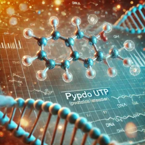
Pseudo UTP (Pseudouridine-5′-triphosphate) is a chemically modified nucleotide that has revolutionized RNA therapeutics by enhancing the stability and functionality of synthetic mRNA. The GMP-grade Pseudo UTP Tris Solution (100 mM) is essential for clinical and industrial applications requiring high-quality RNA synthesis.

This solution is widely used in advanced research and pharmaceutical production, including mRNA-based vaccine development, gene editing, and synthetic biology. With the growing interest in mRNA technology, understanding the significance of pseudo UTP is crucial for researchers, biotech firms, and regulatory bodies.
Understanding Pseudo UTP

Pseudo UTP is a uridine analog where uridine is replaced with pseudouridine, an alternative form of the nucleoside that enhances mRNA stability, translation efficiency, and immunogenicity reduction (NCBI). Researchers at Harvard Medical School and Stanford University have extensively studied pseudouridine’s role in optimizing mRNA-based drugs.

Studies from the Centers for Disease Control and Prevention (CDC) and National Institutes of Health (NIH) highlight how pseudouridine modification reduces immune activation, a key factor in vaccine safety and efficiency.

Key Applications of Pseudo UTP Tris Solution (100 mM)

This highly purified 100 mM GMP-grade solution is indispensable in numerous biotechnological and pharmaceutical applications:

1. mRNA-Based Vaccines

  • Pseudo UTP enhances the performance of mRNA vaccines like those used for COVID-19 (CDC).
  • Reduces unwanted immune responses, ensuring better tolerance and efficiency in humans.

2. Gene Therapy and CRISPR Technologies

  • The use of pseudouridine-modified mRNA in CRISPR/Cas9 gene editing enhances the precision of genetic modifications (NIH).
  • Used in research on genetic disorders, rare diseases, and regenerative medicine (FDA).

3. In Vitro Transcription (IVT) Reactions

  • Pseudo UTP is used in IVT reactions to synthesize high-quality mRNA for research and therapeutic applications (NCBI).
  • Compatible with T7, T3, and SP6 RNA polymerases, ensuring a seamless RNA production process (MIT).

4. Synthetic Biology and RNA Engineering

  • Synthetic biology advancements rely on modified nucleotides like Pseudo UTP for designing functional RNA structures (University of California).
  • Researchers from Johns Hopkins University explore the role of pseudouridine in RNA folding and cellular functions.
Why Choose GMP-Grade Pseudo UTP?

1. High Purity and Consistency

  • GMP-certified manufacturing ensures minimal contamination and batch-to-batch reproducibility (US FDA).
  • Meets stringent requirements for pharmaceutical and clinical research.

2. Enhanced RNA Stability

  • Pseudouridine incorporation increases RNA half-life, making it ideal for long-lasting therapeutic applications (NCBI).

3. Improved Translational Efficiency

  • Modified mRNA synthesized using pseudo UTP has higher translational efficiency in human and mammalian cells (NIH).

4. Regulatory Compliance

  • US FDA regulations ensure high standards for use in human therapeutics (FDA).
  • Adheres to EMA and ICH Guidelines for drug development (European Medicines Agency).
  • ISO 9001 & 13485 certification ensures compliance with international standards (ISO official).

Future Prospects of Pseudo UTP in Biotechnology

As mRNA-based therapies expand into fields such as cancer immunotherapy, autoimmune diseases, and personalized medicine, the demand for GMP-grade Pseudo UTP Tris Solution is expected to increase exponentially.

Advancements in Cancer Research

  • mRNA vaccines targeting cancer cells utilize pseudo UTP-modified mRNA to enhance immune responses against tumors (NCBI).
  • Scientists at Memorial Sloan Kettering Cancer Center are investigating pseudouridine’s role in oncology research.

Expansion of RNA Therapeutics

  • Research institutions like MIT and Harvard predict a breakthrough in RNA-based regenerative medicine.
  • The rise of personalized mRNA therapies will require high-quality modified nucleotides for precision treatments (NIH).

Conclusion

With its superior stability, GMP-grade Pseudo UTP Tris Solution (100 mM) is a game-changer in mRNA-based therapies and biotechnological research. Institutions such as NIH, CDC, FDA, and leading universities continue to invest in developing more advanced RNA-based medical solutions.

For further information, researchers and biotech professionals can explore:

  • NIH – Funding opportunities and research breakthroughs
  • FDA – Regulatory insights for pharmaceutical applications
  • NCBI – Latest scientific publications on pseudouridine and RNA modifications
  • CDC – Updates on RNA-based vaccine development

The future of RNA medicine relies on innovations in nucleotide modifications, and Pseudo UTP Tris Solution (100 mM) remains at the forefront of this evolution.
 

Rat Primary Schwann Cells: Isolation, Function, and Research Applications

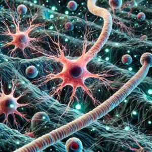
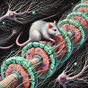
Introduction Schwann cells are essential glial cells in the peripheral nervous system (PNS), responsible for forming the myelin sheath around neuronal axons, which facilitates rapid nerve impulse conduction. These cells are crucial in nerve regeneration and repair, making them a key focus of neurological and regenerative medicine research. Rat primary Schwann cells have been widely used in experimental models to explore cellular mechanisms underlying myelination, neurotrophic support, and nerve regeneration.

This article provides an in-depth overview of rat primary Schwann cells, their isolation methods, biological functions, and research applications.

Isolation and Culture of Rat Primary Schwann Cells

The isolation of primary Schwann cells from rat peripheral nerves is a fundamental step in studying their biology and therapeutic potential. Standard protocols involve the dissection of the sciatic nerve followed by enzymatic digestion to obtain a single-cell suspension. The most common methods include:

  • Pre-degeneration technique: This involves culturing the dissected nerves for several days before enzymatic dissociation, which enhances Schwann cell yield and purity (NIH.gov).
  • Enzymatic digestion: Treatment with enzymes such as collagenase and trypsin helps break down the extracellular matrix, releasing Schwann cells from the nerve tissue (PubMed.gov).
  • Purification techniques: The cultures are often treated with mitotic inhibitors such as cytosine arabinoside to remove contaminating fibroblasts, ensuring high-purity Schwann cell cultures (NCBI.nlm.nih.gov).

Cultured Schwann cells can proliferate in vitro under appropriate conditions, providing a valuable model for studying myelination, cellular interactions, and regenerative processes. Serum-free media optimized for Schwann cells have been developed to maintain their functional integrity (scholars.uky.edu).

Biological Functions of Schwann Cells

Schwann cells play several critical roles in peripheral nerve function and repair. Their primary functions include:

  • Myelination: Schwann cells wrap around axons to form the myelin sheath, which insulates nerve fibers and facilitates rapid signal transmission. Studies show that neuregulin signaling regulates Schwann cell differentiation and myelination (NSF.gov).
  • Axonal support and regeneration: Following nerve injury, Schwann cells dedifferentiate and adopt a repair phenotype that supports axonal regrowth. They secrete neurotrophic factors such as nerve growth factor (NGF) and glial cell-derived neurotrophic factor (GDNF) to promote regeneration (MedlinePlus.gov).
  • Immune modulation: Schwann cells contribute to the immune response in the PNS by interacting with macrophages and modulating inflammatory processes during nerve repair (CDC.gov).

Research Applications of Rat Primary Schwann Cells

1. Peripheral Nerve Regeneration Studies

Rat Schwann cells are widely used in models of nerve injury and repair. Implanting Schwann cell-seeded nerve grafts enhances regeneration in animal models of peripheral nerve injury (NIH RePORTER). Research has also explored using biomaterial scaffolds embedded with Schwann cells to facilitate nerve regeneration (FDA.gov).

2. Myelination and Demyelination Disorders

Experimental models utilizing rat Schwann cells have provided insights into demyelinating diseases such as Charcot-Marie-Tooth disease and Guillain-Barré syndrome. These models help assess potential therapies for myelin repair (HHS.gov).

3. Schwann Cell Transplantation for Spinal Cord Injury

Schwann cell transplantation has been explored as a potential treatment for spinal cord injuries. Studies demonstrate that transplanted Schwann cells can promote axonal growth and remyelination in the injured spinal cord (ClinicalTrials.gov).

4. Neuroinflammation and Neuropathic Pain

Recent research has investigated the role of Schwann cells in neuroinflammatory conditions and chronic pain. Rat Schwann cells are used to study cytokine signaling and pain mechanisms in neuropathic conditions (Johns Hopkins Medicine).

Future Directions and Challenges

While rat Schwann cells provide an invaluable model for peripheral nerve research, several challenges remain:

  • Improving culture methods: Current protocols must be refined to enhance Schwann cell survival and maintain their regenerative phenotype (NIH Blueprint).
  • Enhancing therapeutic applications: Further research is needed to optimize Schwann cell-based therapies for clinical translation (NINDS.nih.gov).
  • Understanding Schwann cell plasticity: Investigating the molecular mechanisms governing Schwann cell dedifferentiation and repair potential may open new avenues for treatment (Harvard.edu).

Conclusion

Rat primary Schwann cells serve as a crucial tool for studying peripheral nerve biology, myelination, and regenerative medicine. Their ability to support axonal growth and modulate the immune response makes them promising candidates for therapeutic interventions in nerve injuries and neurodegenerative disorders.

As research advances, Schwann cell-based strategies could revolutionize treatments for peripheral nerve damage, spinal cord injuries, and demyelinating diseases. By leveraging cutting-edge molecular and cellular techniques, scientists continue to unlock the potential of Schwann cells in neuroscience and regenerative medicine.

Exploring Primary Rabbit Myocardial Cells (MC): Isolation, Applications, and Future Directions

Introduction

Primary rabbit myocardial cells (MCs) are a crucial model for studying cardiovascular physiology, disease mechanisms, and potential therapeutic interventions. Due to their physiological and electrophysiological similarities to human cardiomyocytes, they are widely utilized in translational research. This article delves into the isolation, culture, applications, advantages, challenges, and future directions of primary rabbit myocardial cells, providing valuable insights and references to authoritative sources from government and educational institutions.

Isolation and Culture of Primary Rabbit Myocardial Cells

The process of isolating primary rabbit myocardial cells requires precision and technical expertise. Researchers typically employ enzymatic digestion techniques using collagenase and protease to break down the extracellular matrix and release viable cardiomyocytes. Studies have demonstrated effective isolation protocols that maintain cell viability and functionality for in vitro experiments. Detailed methodologies can be found in resources from the National Institutes of Health (NIH) and the National Center for Biotechnology Information (NCBI).

Once isolated, these cells must be cultured in optimized conditions that support their contractile activity and electrophysiological properties. The media composition, temperature, CO2 concentration, and substrate coating play a critical role in cell survival and function. The Johns Hopkins University School of Medicine provides extensive research on the maintenance of cardiac cells in vitro.

Applications in Cardiovascular Research

Electrophysiology Studies

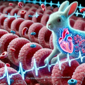
One of the primary applications of primary rabbit myocardial cells is in electrophysiology research. These cells have been instrumental in understanding the properties of ion channels, such as ATP-sensitive potassium (K_ATP) channels, which regulate heart rhythm and cardiac excitability. A study published by the American Heart Association highlights the significance of rabbit myocardial cells in investigating arrhythmias and cardiac conduction disorders.

Heart Failure and Cardiomyopathies

Rabbit myocardial cells are also used to explore the mechanisms underlying heart failure and cardiomyopathies. Research from Harvard Medical School has provided insights into the molecular and cellular changes in failing hearts. Studies on calcium handling abnormalities in rabbit cardiomyocytes have contributed to our understanding of how intracellular calcium dysregulation leads to heart failure, as discussed in NIH’s National Heart, Lung, and Blood Institute (NHLBI) resources.

Drug Testing and Toxicology

Another crucial application of rabbit myocardial cells is in pharmacological research and drug testing. The cells serve as an effective platform for evaluating the cardiotoxic effects of new drugs before clinical trials. Regulatory agencies like the U.S. Food and Drug Administration (FDA) emphasize the importance of preclinical cardiac toxicity assessments using relevant animal models, including rabbit-derived myocardial cells.

Advantages of Using Rabbit Myocardial Cells

Physiological Similarities to Human Hearts

Rabbit hearts exhibit electrophysiological properties similar to those of human hearts, including comparable action potential durations and ion channel expression. This makes them preferable over smaller rodent models such as mice and rats, as noted in studies from the National Library of Medicine (NLM).

Larger Cell Size for Manipulation

Compared to smaller rodent models, rabbit cardiomyocytes are easier to manipulate due to their larger size. This facilitates more precise experimental interventions, such as patch-clamp recordings and genetic modifications, as discussed in research from the University of California, San Francisco (UCSF).

Challenges and Ethical Considerations

While primary rabbit myocardial cells offer many advantages, their use also comes with challenges. The isolation process is technically demanding and can result in low yields if not performed correctly. Moreover, maintaining these cells in long-term culture while preserving their contractile properties remains a challenge. Researchers at Stanford University have explored novel techniques to improve cell survival and functionality in vitro.

Ethical considerations regarding the use of animals in research must also be addressed. Guidelines from the U.S. Department of Agriculture (USDA) and the Office of Laboratory Animal Welfare (OLAW) outline best practices for the humane treatment of research animals, ensuring compliance with ethical standards.

Recent Advances and Future Directions

Induced Pluripotent Stem Cells (iPSCs)

The advent of induced pluripotent stem cell (iPSC) technology presents an exciting opportunity to generate cardiomyocytes without the need for primary animal cell isolation. Researchers at the University of Pennsylvania are actively working on deriving rabbit-specific iPSC cardiomyocytes to replace traditional primary cell models.

Gene Editing and CRISPR Applications

CRISPR-Cas9 gene-editing technology is being increasingly applied to rabbit models to study the genetic underpinnings of cardiovascular diseases. Institutions such as the Massachusetts Institute of Technology (MIT) and NIH are pioneering research on CRISPR-mediated modifications in cardiac cells to investigate genetic contributions to heart disease.

3D Bioprinting of Cardiac Tissue

Recent advancements in 3D bioprinting are paving the way for engineering cardiac tissue constructs using rabbit myocardial cells. Research from the University of Michigan suggests that bioprinted cardiac tissues could serve as superior models for drug testing and regenerative medicine.

Conclusion

Primary rabbit myocardial cells continue to play a pivotal role in cardiovascular research, offering unparalleled insights into heart function, disease mechanisms, and therapeutic development. Despite challenges, advancements in cell culture techniques, gene editing, and tissue engineering are set to enhance their utility. As research evolves, collaborations between government agencies, academic institutions, and biotech companies will further drive innovation in cardiac science.

For more information on myocardial cell research, visit reputable sources such as the National Institutes of Health (NIH), the Centers for Disease Control and Prevention (CDC), and the World Health Organization (WHO).