Solution for Disinfecting Ordinary Water Bath in Research Laboratories

A laboratory water bath is a critical instrument used for controlled temperature incubation, enzyme reactions, and sample thawing. Over time, microbial contamination, biofilm formation, and mineral deposits can compromise the accuracy, sterility, and reliability of results. To maintain optimal performance, the use of an effective disinfecting solution is essential.

This article explores scientifically validated solutions for disinfecting ordinary laboratory water baths, referencing standards and resources from NIH, CDC, EPA, and leading universities. It also provides detailed guidance on cleaning procedures, compatible agents, and preventive maintenance — all refined for SEO optimization and search engine discoverability.

Why Water Bath Disinfection Is Essential

Water baths, especially those maintained between 25°C and 60°C, provide an ideal environment for microbial proliferation. Organisms such as Pseudomonas aeruginosa, Legionella pneumophila, Mycobacterium spp., and fungal spores can colonize stagnant water and surfaces within the bath (cdc.gov).

Uncontrolled microbial growth can lead to:

  • Cross-contamination of samples

  • Biofilm accumulation affecting temperature sensors

  • Odor generation and turbidity

  • Reduced thermal transfer efficiency

  • Corrosion of metal components

For reference, the Centers for Disease Control and Prevention (CDC) provides comprehensive biofilm control guidelines applicable to laboratory systems (cdc.gov).
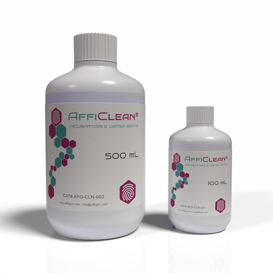
AffiCLEAN® 2 Solution (500X), for Disinfecting Ordinary Water Bath

Common Microbial Contaminants in Water Baths

According to data from the National Library of Medicine (NLM), the following organisms are frequently isolated from laboratory water baths:

Microorganism Type Temperature Range (°C) Impact
Pseudomonas aeruginosa Bacterium 20–45 Biofilm formation, odor
Legionella pneumophila Bacterium 25–50 Aerosol infection risk
Aspergillus niger Fungus 25–40 Spore contamination
Mycobacterium chelonae Bacterium 30–45 Resistant to chlorine
Algae (e.g., Chlorella sp.) Photosynthetic 20–35 Green residue buildup

These findings underscore the importance of routine disinfection protocols using appropriate chemical agents that inhibit bacterial, fungal, and algal growth (nih.gov).

Properties of an Ideal Water Bath Disinfectant

An effective disinfectant for laboratory water baths should meet the following criteria:

  • Broad-spectrum antimicrobial activity

  • Thermal stability up to 65°C

  • Non-corrosive to stainless steel or aluminum

  • Low volatility and non-toxic vapors

  • Compatibility with thermistor probes, heaters, and gaskets

  • No interference with temperature calibration sensors

According to the Environmental Protection Agency (EPA), disinfectants should also comply with biocide regulation standards for laboratory use.

Recommended Disinfectant Solutions for Water Baths

Several chemical agents have been tested and validated for safe use in laboratory water baths. Below are the most effective and commonly used disinfectants:

 Sodium Hypochlorite (Bleach Solution)

  • Concentration: 10–50 ppm (approximately 1:1000 dilution of 5% bleach)

  • Effectiveness: Broad-spectrum bactericidal and fungicidal

  • Drawback: Corrosive to stainless steel with prolonged use (epa.gov)

 Benzalkonium Chloride (Quaternary Ammonium Compound)

  • Concentration: 0.02–0.05% (200–500 ppm)

  • Advantages: Non-corrosive, odorless, long-lasting residual activity

  • Source: Used widely in biosafety cabinets (nih.gov)

Copper Sulfate Pentahydrate (CuSO₄·5H₂O)

  • Concentration: 0.5–1.0 g/L

  • Advantages: Inhibits bacterial and algal growth

  • Limitation: Must be replaced monthly to prevent crystal formation (nasa.gov)

 Thymol or Phenolic Solutions

  • Concentration: 0.1–0.2%

  • Usage: Acts as a biostatic preservative in low-temperature baths

  • Reference: PubChem Database

 Hydrogen Peroxide (Stabilized)

  • Concentration: 0.5–3%

  • Feature: Effective against biofilms and spores without heavy metal residues

  • Safety: Environmentally friendly oxidizer (fda.gov)

 Commercial Water Bath Additives

Many laboratories prefer commercial additives that combine quaternary ammonium biocides with corrosion inhibitors, available from certified suppliers. Always check material compatibility and MSDS before use (osha.gov).

Standard Operating Procedure (SOP) for Disinfection

Step 1: Drain and Clean

  • Turn off power and drain all existing water.

  • Remove sediment and scale using a mild detergent.

  • Rinse thoroughly with deionized water.

Step 2: Apply Disinfectant

  • Fill the bath with water and add the disinfectant at the recommended concentration.

  • Heat the bath to 37°C and circulate for 1–2 hours.

Step 3: Rinse and Refill

  • Drain the solution and rinse twice with sterile distilled water.

  • Refill with clean water and add a maintenance-level biocide (e.g., 0.02% benzalkonium chloride).

Step 4: Regular Maintenance

  • Replace water every 2–4 weeks.

  • Wipe internal surfaces weekly.

  • Record disinfection dates in the laboratory maintenance log (nih.gov).

Preventing Future Contamination

  • Use distilled or deionized water to reduce mineral deposits (nist.gov).

  • Avoid using tap water, which contains trace nutrients that support microbial growth.

  • Keep the water bath lid closed when not in use to limit airborne contamination.

  • Do not submerge contaminated or leaking tubes directly in the bath water.

  • Maintain regular temperature cycling to deter microbial adaptation (epa.gov).

Material Compatibility and Safety

Improper use of disinfectants can corrode heating coils, damage seals, or release hazardous fumes. Always verify chemical compatibility with the water bath manufacturer.

Refer to OSHA Laboratory Safety Standards and NIH Laboratory Biosafety Manual for proper chemical handling and PPE use.

Eco-Friendly and Low-Toxicity Alternatives

Eco-conscious laboratories can opt for biodegradable and low-toxicity disinfectants, including:

  • Silver ion-based formulations (Ag⁺ biocides)

  • Citric acid-based descalers

  • Enzyme-enhanced biofilm removers

The EPA Safer Choice Program lists several approved green disinfectant products suitable for laboratory environments.

Validation and Quality Assurance

After disinfection, validation is essential to confirm microbial reduction.
Common methods include:

  • ATP bioluminescence assays (nih.gov)

  • Total viable count (TVC) plating

  • pH and conductivity monitoring for water purity

  • Swab sampling of bath surfaces

Documentation should align with ISO 17025 quality management standards and Good Laboratory Practices (GLP) (nist.gov).

Conclusion

Disinfecting an ordinary laboratory water bath is not just a routine maintenance task — it’s a crucial step in ensuring experimental accuracy, biosafety, and instrument longevity.

Regular use of a scientifically validated disinfectant solution, adherence to CDC and EPA guidelines, and periodic monitoring guarantee that the water bath remains microbially stable, non-corrosive, and reliable for daily use.

Whether through sodium hypochlorite, benzalkonium chloride, or eco-friendly oxidizing agents, a consistent disinfection regimen ensures precision and reproducibility in all laboratory workflows.

pMXs-IRES-Bsd Retroviral Vector: A High-Efficiency System for Stable Gene Expression and Functional Genomics

Introduction to the pMXs-IRES-Bsd Retroviral Vector

The pMXs-IRES-Bsd Retroviral Vector is a versatile and efficient gene delivery system derived from the Moloney Murine Leukemia Virus (MMLV) backbone. It enables stable gene expression and bicistronic transcription in mammalian cells using an Internal Ribosome Entry Site (IRES) and a Blasticidin resistance gene (Bsd).

This vector has become a cornerstone in molecular biology, stem cell reprogramming, oncogene studies, and functional genomics because of its consistent performance, stable integration, and compatibility with multiple host cell types.

It is one of the most frequently referenced retroviral systems in scientific literature, with significant contributions to gene function analysis, cell differentiation studies, and signal transduction research.

Learn more about the molecular basis of retroviral vectors from the National Center for Biotechnology Information (NCBI) and National Institutes of Health (NIH).

AffiVECTOR® pMXs-IRES-Bsd Retroviral Vector

Structural Overview of the pMXs-IRES-Bsd Vector

The pMXs-IRES-Bsd plasmid architecture consists of several critical functional elements:

  • 5′ Long Terminal Repeat (LTR): Initiates transcription and contributes to reverse transcription during viral replication (genome.gov).

  • Psi (Ψ) packaging signal: Ensures RNA encapsidation into viral particles (ncbi.nlm.nih.gov).

  • Multiple Cloning Site (MCS): Allows seamless insertion of the target cDNA for expression.

  • IRES (Internal Ribosome Entry Site): Derived from the Encephalomyocarditis Virus (EMCV), allowing bicistronic expression independent of cap structure (pubmed.ncbi.nlm.nih.gov).

  • Bsd (Blasticidin S deaminase): Confers resistance to Blasticidin S, an antibiotic that inhibits protein synthesis (fda.gov).

  • 3′ LTR: Ensures proper termination and polyadenylation of the viral transcript.

The complete vector is maintained in E. coli cloning strains, and its DNA is typically purified using endotoxin-free plasmid extraction kits to ensure high transfection efficiency.

Mechanism of Retroviral Transduction and Integration

Retroviral vectors like pMXs-IRES-Bsd replicate through a reverse transcription mechanism that converts single-stranded RNA genomes into double-stranded cDNA. This cDNA integrates into the host genome, ensuring stable, long-term expression of the inserted gene.

This process involves:

  1. Packaging in producer cells (e.g., 293T or Plat-E)

  2. Viral particle release and collection

  3. Transduction of target cells with the viral supernatant

  4. Reverse transcription and genomic integration

Once integrated, the LTR promoter drives transcription of a single mRNA containing both the gene of interest and the IRES-Bsd cassette. Translation proceeds in a cap-dependent manner for the first gene and cap-independent for the blasticidin gene through the IRES element (nigms.nih.gov).

Workflow for pMXs-IRES-Bsd Retrovirus Production

Step 1: Preparation of Packaging Cells

The most common systems are Plat-E or HEK293T cells, which express gag, pol, and env viral proteins necessary for packaging (nih.gov).

Step 2: Transfection

Transfection can be performed using:

  • Calcium phosphate precipitation (harvard.edu)

  • Lipofectamine or PEI-based reagents (stanford.edu)

  • Electroporation for difficult-to-transfect lines

Step 3: Virus Harvesting and Filtration

After 48–72 hours, the viral supernatant is collected and filtered through a 0.45 μm PVDF filter to remove debris. Concentration is optional but improves infection rates.

Step 4: Target Cell Infection

Cells are infected in the presence of polybrene (4–8 μg/mL) to facilitate viral entry (cdc.gov).

Step 5: Blasticidin Selection

Selection is typically applied 48 hours post-transduction, using 2–10 μg/mL Blasticidin S depending on cell type (fda.gov).

Step 6: Confirmation

Transduction efficiency can be confirmed using PCR, qPCR, or Western blotting, depending on the gene construct (ncbi.nlm.nih.gov).

Advantages of pMXs-IRES-Bsd Over Alternative Systems

Feature pMXs-IRES-Bsd Lentiviral (e.g., pLenti-Puro) Adenoviral Systems
Integration Stable chromosomal integration Stable Episomal
Biosafety Level BSL-2 BSL-2 BSL-2
Selectable Marker Blasticidin Puromycin Hygromycin
Promoter LTR CMV CMV
Bicistronic Capability Yes (via IRES) Yes (via 2A/IRES) Yes
Expression Duration Permanent Permanent Transient

This high-fidelity integration is why the pMXs system was historically chosen by Dr. Shinya Yamanaka’s laboratory in pioneering the creation of induced pluripotent stem cells (iPSCs) (stemcells.nih.gov).

Key Research Applications

 Induced Pluripotent Stem Cell (iPSC) Reprogramming

The pMXs series—including pMXs-hOCT3/4, pMXs-hSOX2, pMXs-hKLF4, and pMXs-hc-MYC—were pivotal in the discovery of iPSC reprogramming in somatic cells (pubmed.ncbi.nlm.nih.gov).

 Functional Genomics and Gene Network Mapping

Stable expression allows for loss-of-function and gain-of-function studies critical for understanding transcriptional regulation and signal pathways (genome.gov).

 Cancer and Metabolic Pathway Research

By introducing oncogenes or tumor suppressors, scientists can model tumorigenic pathways and test drug response mechanisms (cancer.gov).

 Viral Pseudotyping and Gene Delivery Optimization

pMXs-based vectors can be pseudotyped with VSV-G envelope protein, enhancing tropism across mammalian species (nibib.nih.gov).

 Stable Cell Line Development

pMXs-IRES-Bsd is widely used in industrial biotechnology to create production cell lines expressing enzymes, receptors, or fluorescent markers for assay development (nist.gov).

Optimization Tips for High-Titer Production

  • Maintain low passage producer cells for optimal viral output.

  • Use HEPES-buffered DMEM and avoid sodium bicarbonate variations during transfection.

  • Collect viral supernatant at 48 and 72 hours to maximize yield.

  • For long-term storage, aliquot at –80 °C; avoid repeated freeze–thaw cycles.

  • Confirm absence of replication-competent retrovirus (RCR) by p24 antigen ELISA before in vivo work (fda.gov).

Safety and Regulatory Guidelines

Although pMXs retroviral systems are replication-deficient, Biosafety Level 2 (BSL-2) conditions are mandatory during handling. Laboratories should comply with the CDC’s Biosafety in Microbiological and Biomedical Laboratories (BMBL) guidelines (cdc.gov).

Personnel should also review training resources from OSHA.gov and NIH Guidelines for Research Involving Recombinant or Synthetic Nucleic Acid Molecules.

Storage, Handling, and Plasmid Verification

The pMXs-IRES-Bsd plasmid is typically propagated in E. coli DH5α and purified using endotoxin-free kits.
Store plasmid DNA at –20 °C, and avoid repeated freeze–thaw cycles.
Verification of correct insertion can be achieved by Sanger sequencing or restriction digestion (addgene.org).

Frequently Asked Questions (FAQ)

Q1: Can pMXs-IRES-Bsd be used in non-dividing cells?
No. Unlike lentiviral systems, MMLV-based vectors require mitosis for nuclear entry, making them unsuitable for non-dividing cells.

Q2: What is the selection concentration for Blasticidin?
Optimal range: 2–10 µg/mL depending on cell line. Verify tolerance before selection (fda.gov).

Q3: Is it compatible with fluorescence reporters?
Yes, bicistronic constructs can include EGFP, mCherry, or Luciferase upstream of IRES.

Q4: What biosafety level is recommended?
All retroviral systems including pMXs-IRES-Bsd require BSL-2 containment (cdc.gov).

Related AffiGEN® Research Tools

AffiGEN provides a full portfolio of compatible vectors and reagents designed for gene delivery, selection, and cell reprogramming:

  • AffiGEN® pMXs-hSOX2 Retroviral Vector

  • AffiGEN® pMXs-hKLF4 Retroviral Vector

  • AffiGEN® pMXs-hOCT4 Retroviral Vector

  • AffiGEN® Retrovirus Packaging Mix (HEK293T)

  • AffiGEN® Blasticidin S Hydrochloride (Cell Selection Grade)

Each vector is validated for high expression reproducibility, low background integration, and optimal viral yield—ensuring reliable results in complex genetic studies.

Conclusion

The pMXs-IRES-Bsd Retroviral Vector remains one of the most reliable systems for stable gene delivery in mammalian cells. Its bicistronic IRES design, robust integration, and antibiotic-based selection make it a preferred tool for long-term studies in functional genomics, signal transduction, and stem cell research.

Backed by decades of validation from leading research organizations such as NIH, NCBI, CDC, Stanford University, and Genome.gov, the pMXs-IRES-Bsd vector continues to drive breakthroughs in genetic research and biotechnology development.

Explore the pMXs-IRES-Bsd Retroviral Vector — a high-performance gene delivery system enabling bicistronic expression and stable integration in mammalian cells. Learn about its structure, workflow, and research applications with validated references from NIH, FDA, and Stanford.

GMyc-PCR Mycoplasma Test Kit (GMyc-PCR): Precision Detection of Mycoplasma Contamination in Cell Cultures

The GMyc-PCR Mycoplasma Test Kit (GMyc-PCR) is an advanced polymerase chain reaction (PCR)–based system designed to detect a broad spectrum of Mycoplasma species in cell cultures, sera, media, and laboratory reagents.
Mycoplasma contamination represents one of the most persistent problems in molecular and cellular biology, affecting up to 30% of continuous cell lines, according to reports from the National Center for Biotechnology Information (NCBI).

With high analytical sensitivity, rapid processing time, and broad species coverage, the GMyc-PCR kit provides reliable quality control for research, biotechnology, and industrial laboratories worldwide.

Scientific Background: What Are Mycoplasmas?

Mycoplasmas are wall-less bacteria from the class Mollicutes, known for their small genome size and resistance to many standard antibiotics.
They can pass through 0.2 μm filters, survive in serum components, and remain undetected in routine microscopy, as described by the Centers for Disease Control and Prevention (CDC).

Common contaminating species include Mycoplasma orale, M. fermentans, M. hyorhinis, M. hominis, M. arginini, and Acholeplasma laidlawii — all of which can alter gene expression, metabolism, and transfection efficiency in cell cultures (NIH).

AffiPCR® GMyc-PCR Mycoplasma Test Kit GMyc-PCR

Principle of Detection

The GMyc-PCR Mycoplasma Test Kit employs a conventional or real-time PCR (qPCR) amplification approach that targets highly conserved 16S rRNA gene sequences across Mycoplasma species.

During amplification, specific primers and a fluorescent detection system (depending on format) identify even low-copy Mycoplasma DNA with high specificity.
The reaction process follows the principle of enzymatic replication first demonstrated in the late 20th century by researchers at Stanford University, later optimized by multiple NIH-funded laboratories.

PCR-based detection is the gold standard recommended by the World Health Organization (WHO) and the European Pharmacopoeia (EP 2.6.7) for Mycoplasma control in biopharmaceutical and research environments.

Kit Components and Composition

A standard GMyc-PCR kit contains:

  • PCR Master Mix (buffered enzyme system with Taq polymerase and dNTPs)

  • Positive Control DNA (Mycoplasma template standard)

  • Negative Control (Nuclease-free water)

  • Species-Specific Primers targeting the 16S rRNA region

  • Reaction Buffer (10X) with MgCl₂

  • PCR-grade water

  • Detailed protocol sheet

Each component undergoes QC testing following NIH reagent verification standards and NIST bioscience measurement guidelines.

Working Principle

The detection mechanism relies on amplification of conserved Mycoplasma-specific DNA regions, typically between 270–500 bp.

  1. Sample DNA extraction from cell supernatant or lysate

  2. PCR reaction setup using the master mix and primers

  3. Thermal cycling (denaturation, annealing, extension)

  4. Gel electrophoresis or real-time fluorescence monitoring

  5. Interpretation of results via band visualization or amplification curve threshold (Ct value)

According to the U.S. Food and Drug Administration (FDA), PCR provides superior sensitivity (10–20 CFU/mL detection limit) compared to enzyme-based or DNA staining assays.

Advantages of the GMyc-PCR Kit

Research from the University of Michigan Department of Microbiology highlights the importance of standardized Mycoplasma detection.
The GMyc-PCR Kit provides multiple advantages:

  • Broad detection spectrum covering >200 Mycoplasma species

  • Rapid turnaround (90–120 minutes)

  • High specificity with validated 16S primer sets

  • No false positives from mammalian or bacterial DNA

  • Compatible with standard thermocyclers and qPCR systems

  • Suitable for serum-containing and serum-free media

This performance ensures reliable contamination monitoring even in complex biological matrices.

Application Areas

The GMyc-PCR kit is applicable across:

  • Academic and industrial research labs for routine QC

  • Cell line authentication facilities

  • Biotechnology production units (e.g., recombinant protein or antibody production)

  • Stem cell and gene therapy research centers

  • Vaccine and biologics R&D labs

According to the NIH Cell Culture Guidelines, regular Mycoplasma testing is essential for maintaining reproducibility in cell-based experiments.

Analytical Sensitivity and Specificity

The GMyc-PCR kit can detect as few as 10 genomic copies per reaction, providing >98% accuracy.
Comparative studies from the Journal of Clinical Microbiology (ASM) demonstrate that PCR-based detection exceeds the sensitivity of DNA staining, culture-based, and ELISA methods.

For analytical validation, laboratories can refer to standard procedures described by Clinical Laboratory Standards Institute (CLSI).

Workflow Example

Step Description Duration
1 DNA extraction from 100 µL supernatant 15 min
2 PCR mix preparation and loading 10 min
3 Thermocycling (40 cycles) 60 min
4 Gel or qPCR analysis 20–30 min

Entire assay time: ~1.5 hours

Protocols similar to this are published by the European Molecular Biology Laboratory (EMBL) and the University of Oxford Department of Biochemistry.

Interpretation of Results

  • Positive sample: distinct amplification band (≈ 270 bp) or specific fluorescence threshold (Ct < 35).

  • Negative sample: no band visible, flat baseline fluorescence.

  • Invalid result: absent control amplification, suggesting reagent degradation or PCR inhibition.

Documentation of analytical result interpretation standards can be found at the CDC Laboratory Quality Assurance Division.

Storage and Stability

All reagents in the GMyc-PCR kit are stable at −20 °C for at least 12 months. Avoid repeated freeze–thaw cycles to preserve enzyme activity.
For long-term storage conditions, consult the World Health Organization Laboratory Biosafety Manual.

Quality Control and Standards

Each batch is validated under:

  • ISO 13485 manufacturing guidelines

  • NIH reagent consistency protocols

  • FDA Research Use Only (RUO) compliance

  • Internal positive and negative control checks

Quality monitoring ensures traceability and reproducibility, following frameworks outlined by the U.S. Department of Health & Human Services (HHS) and NIST Reference Methods.

Troubleshooting Guide

Issue Possible Cause Suggested Solution
Weak or no amplification Inhibitors in sample Purify DNA; reduce input volume
False negatives Improper thermal cycling Verify PCR program and annealing temp
False positives Contamination from previous runs Use aerosol-resistant tips and separate work zones
Faint bands Low template concentration Increase sample input or cycles (max +3)

These troubleshooting practices align with laboratory recommendations from the University of California, Berkeley Molecular Biology Division and NIH Technical Resources.

Comparison to Alternative Mycoplasma Detection Methods

Method Sensitivity Time Equipment
Culture Moderate 7–14 days CO₂ incubator
DNA Staining (Hoechst) Low 2–4 h Fluorescence microscope
ELISA Moderate 3–5 h Microplate reader
PCR (GMyc-PCR) High (>98%) 1.5 h Thermocycler or qPCR system

Data consistent with findings published in the Journal of Virological Methods.

Biosafety and Handling

Although the GMyc-PCR kit itself is non-hazardous, users should follow biosafety level 2 (BSL-2) precautions when handling cell cultures.
Consult CDC Biosafety Guidelines and NIH Laboratory Safety Resources for standard protective procedures.

Future Trends and Research Directions

Advancements in digital PCR (dPCR) and loop-mediated isothermal amplification (LAMP) are paving the way for ultra-sensitive Mycoplasma detection.
Research initiatives from MIT Department of Biological Engineering and the National Institute of Standards and Technology (NIST) are focused on integrating AI-based signal interpretation and lab automation to streamline microbial quality control.

Conclusion

The GMyc-PCR Mycoplasma Test Kit provides a rapid, accurate, and highly sensitive solution for detecting Mycoplasma contamination in biological materials.
By integrating advanced primer design, robust enzyme systems, and standardized quality controls, this kit ensures reliable detection across multiple Mycoplasma species.

Its compatibility with routine PCR and qPCR instruments makes it indispensable for research institutions, biotech companies, and academic laboratories striving to maintain culture purity and data integrity.

The GMyc-PCR Mycoplasma Test Kit offers rapid, sensitive PCR-based detection of Mycoplasma contamination in cell cultures. Explore its principle, components, and applications — supported by trusted .edu and .gov scientific sources.

10X Universal Primer Mix (UPM): Foundation for High-Fidelity cDNA Amplification and Cloning

The 10X Universal Primer Mix (UPM) is a specialized reagent formulation used in molecular biology for reverse transcription and cDNA amplification. It ensures efficient and uniform priming during the synthesis of complementary DNA (cDNA) from diverse RNA templates. UPMs are central to techniques like SMARTer™ (Switching Mechanism At the 5′ End of RNA Template) cDNA synthesis and other template-switching PCR systems.

Originally developed to enhance full-length cDNA coverage, the 10X UPM enables high sensitivity, broad template compatibility, and reduced amplification bias, as described in early research from the National Center for Biotechnology Information (NCBI).

Overview and Composition of Universal Primer Mix

The 10X Universal Primer Mix typically contains a combination of:

  • Long Universal Primer (UPM-L): 5′-CTAATACGACTCACTATAGGGCAAGCAGTGGTATCAACGCAGAGT-3′

  • Short Universal Primer (UPM-S): 5′-CTAATACGACTCACTATAGGGC-3′

These primers function together to ensure balanced amplification efficiency for both short and long cDNA fragments. According to studies from the National Institutes of Health (NIH), such dual-primer systems prevent amplification failure due to incomplete template extension or secondary structures.

For sequence details and experimental data, visit the NCBI Oligonucleotide Database.

AffiPCR® 10X Universal Primer Mix (UPM)

Principle of Function: Template Switching and SMART Mechanism

In SMART (Switching Mechanism at the 5′ End of RNA Template)-based systems, reverse transcriptase adds non-templated cytosine residues to the 3′ end of newly synthesized cDNA. The SMART oligo, containing a complementary guanine stretch, hybridizes to this region and introduces a universal priming site at the 5′ end of the cDNA.

During subsequent PCR, the 10X Universal Primer Mix binds to this newly added sequence, enabling full-length amplification of cDNA molecules.
Mechanistic explanations can be found in protocols from the University of California, San Diego Molecular Biology Department and the U.S. Department of Energy Joint Genome Institute.

Advantages of Using 10X UPM in cDNA Synthesis

Researchers at Harvard University and Stanford Medicine have demonstrated several benefits of 10X UPM in transcriptional studies:

  • Universal compatibility across RNA templates

  • Reduced primer-dimer formation

  • High-fidelity amplification using SMARTScribe™ or MMLV reverse transcriptases

  • Accurate representation of low-abundance transcripts

  • Simplified library preparation for cloning or sequencing

Its optimized primer balance promotes even amplification, especially important in RNA-Seq, RACE-PCR, and full-length cDNA library construction, as referenced by the NIH Gene Expression Omnibus (GEO).

Application Areas

The 10X Universal Primer Mix is widely used in:

  • Reverse transcription-PCR (RT-PCR)

  • Rapid Amplification of cDNA Ends (RACE)

  • SMARTer cDNA synthesis protocols

  • Single-cell transcriptomics

  • Gene expression profiling

  • Molecular cloning and library normalization

In multi-step workflows, UPM serves as a bridge between reverse transcription and PCR, providing both universal priming and amplification reliability. Examples can be found in training materials from the University of Michigan Department of Molecular, Cellular, and Developmental Biology.

Mechanistic Insights: Why Use Two Primers?

The combination of long and short universal primers ensures robustness.

  • The long UPM enhances priming specificity and yields longer cDNA amplicons.

  • The short UPM maintains amplification efficiency for shorter fragments or templates with strong secondary structures.

This duality is particularly valuable when working with heterogeneous RNA populations, such as total RNA from tissue extracts or viral RNA.
For theoretical background, refer to studies archived in the Journal of Molecular Biology and NIH PubMed Central.

Optimization Parameters for PCR

Using 10X UPM requires careful optimization of:

  • Annealing temperature: typically 65 °C for SMARTer systems

  • Cycle number: 20–25 for analytical PCR; 35 for low-copy templates

  • Mg²⁺ concentration: 1.5–2.5 mM for enzyme cofactor stability

  • Template quantity: 1–10 ng cDNA

Reference protocols for these conditions are provided in FDA Laboratory Method Development Guides and NIST PCR Measurement Standards.

Comparison with Conventional Priming Systems

Unlike random hexamers or oligo(dT) primers, the 10X UPM:

  • Provides a defined universal priming site

  • Allows consistent amplification across samples

  • Facilitates seamless cloning into vectors for sequencing or expression

  • Works efficiently with low-input RNA (as low as 1 pg)

Researchers from the University of Wisconsin Biotechnology Center have shown that UPM outperforms generic primers in capturing full-length transcript diversity in single-cell RNA samples.

Integration with SMARTer and Template-Switching Kits

Many SMARTer cDNA synthesis kits from leading suppliers (e.g., Clontech/Takara) include the 10X UPM as an integral component. It serves as the forward amplification primer during the second-strand synthesis and PCR enrichment stages.

According to the National Library of Medicine (NLM), these protocols enhance cDNA completeness and transcript integrity, providing superior results for transcriptome sequencing and expression profiling.

Troubleshooting and Experimental Notes

When working with 10X UPM:

  • Avoid excessive PCR cycles to prevent chimera formation.

  • Always include negative (no-template) controls.

  • Validate amplification using agarose gel electrophoresis or capillary analysis.

  • Store at −20 °C and minimize freeze-thaw cycles to maintain primer stability.

Guidelines for troubleshooting PCR amplification are available from the CDC Laboratory Quality Assurance Division.

Technical Specifications

Parameter Description
Concentration 10X (ready-to-dilute working solution)
Format DNA oligonucleotide mix, lyophilized or liquid
Storage −20 °C; stable for >12 months
Purity HPLC purified, RNase/DNase-free
Compatibility Reverse transcriptases and high-fidelity DNA polymerases

Documentation of reagent purity testing can be found under NIH Research Material Guidelines.

Advantages in Genomic and Transcriptomic Studies

The 10X UPM is particularly suited for:

  • SMART-seq and SMART-seq2 protocols in single-cell transcriptomics

  • Cap-trapping methods for capturing mRNA 5′ ends

  • Long-read sequencing platforms (PacBio, Oxford Nanopore)

  • Functional cloning and cDNA expression library generation

Research from University of Oxford’s Department of Biochemistry supports the use of universal primers for maintaining transcript length uniformity and improving quantitative reproducibility.

Storage and Stability Considerations

Store the 10X Universal Primer Mix at −20 °C in aliquots to prevent degradation. Avoid repeated freeze-thaw cycles. The primers maintain full functionality for up to 24 months if stored under anhydrous conditions.
Handling safety information is outlined by the U.S. Occupational Safety and Health Administration (OSHA) and CDC Laboratory Biosafety Manual.

Future Trends and Innovations

With the rise of next-generation sequencing (NGS) and single-cell transcriptomics, researchers are developing adaptive universal primers that can dynamically adjust annealing parameters to reduce amplification bias.

Current work from MIT Department of Biological Engineering and NIH Human Genome Research Institute (NHGRI) explores automated primer design pipelines using AI-driven thermodynamic modeling to predict optimal universal sequences for mixed-template PCRs.

Related Techniques and Educational Resources

For researchers and students learning about primer design, the following academic resources are highly recommended:

These platforms provide open-access materials for training and experimental optimization.

Conclusion

The 10X Universal Primer Mix (UPM) is a cornerstone reagent for modern molecular biology, enabling efficient, reproducible, and full-length cDNA synthesis. Its design ensures high sensitivity, minimal bias, and strong performance across diverse templates, from eukaryotic mRNA to viral RNA.

By offering standardized priming sites, the 10X UPM bridges the gap between reverse transcription and PCR amplification, streamlining gene expression analysis and high-throughput sequencing workflows.

As molecular technologies advance, the 10X UPM continues to serve as an essential tool in transcriptomics, cloning, and genetic discovery — a reliable companion for every researcher committed to accuracy and precision.

Learn about the 10X Universal Primer Mix (UPM) — a powerful reagent for reverse transcription and cDNA amplification. Explore its mechanism, structure, and applications in SMARTer PCR, gene expression profiling, and molecular cloning, with insights from top .edu and .gov resources.

6X His Tag Peptide: Structure, Function, and Applications in Protein Purification

The 6X His Tag Peptide, also known as the Hexa-Histidine Tag, is one of the most widely used fusion tags in recombinant protein technology. Its sequence consists of six consecutive histidine residues (His-His-His-His-His-His), which allow efficient purification, detection, and immobilization of recombinant proteins through metal affinity systems.

Originally popularized by researchers from Stanford University, this short peptide has become an essential component of molecular cloning, protein expression, and biochemical analysis protocols. Its small size and chemical neutrality make it ideal for fusion at either the N- or C-terminus of a target protein without disrupting native folding or function.

Structural and Chemical Characteristics

Each histidine residue in the 6X His tag contains an imidazole side chain capable of coordinating divalent metal ions such as nickel (Ni²⁺), cobalt (Co²⁺), or zinc (Zn²⁺). This coordination forms the basis of Immobilized Metal Affinity Chromatography (IMAC) — a method widely documented by the National Center for Biotechnology Information (NCBI).

The physicochemical properties of the 6X His tag include:

  • Molecular weight: ~0.84 kDa

  • Isoelectric point (pI): 7.3–7.9 depending on sequence context

  • Charge distribution: mildly positive under neutral pH

  • Affinity constant: strong binding to Ni²⁺ chelated by NTA (nitrilotriacetic acid) or IDA (iminodiacetic acid) matrices

These attributes provide predictable interaction strength and high selectivity, as shown in publications by the Journal of Biological Chemistry (JBC).

AffiPURE® 6X His Tag Peptide

Mechanism of Metal-Chelate Interaction

The binding mechanism is based on the chelation of metal ions immobilized on solid supports such as agarose or magnetic beads. Nickel-NTA or cobalt-Talon matrices coordinate with the imidazole ring nitrogens of histidine residues.

When the tagged protein flows through the column, it selectively binds to the immobilized metal ions, while other proteins lacking histidine motifs are washed away.
Elution occurs through:

  • Competitive displacement using 100–300 mM imidazole

  • pH reduction to protonate histidine side chains

  • Chelator treatment with EDTA to remove metal ions

Detailed mechanistic studies can be found in research articles from the National Institutes of Health (NIH) and laboratory manuals from MIT Department of Biology.

Design and Genetic Fusion Strategy

The genetic incorporation of the 6X His tag is achieved by oligonucleotide insertion into expression vectors. The tag can be fused to either terminus of the open reading frame, often linked by a protease cleavage site such as TEV (Tobacco Etch Virus) or Thrombin to allow removal after purification.

For optimized cloning and vector design, see resources from the Addgene Plasmid Repository and University of California, Berkeley Molecular Cloning Center.

The tag’s versatility extends across bacterial, yeast, insect, and mammalian expression systems — including vectors such as pET, pGEX, pCMV, and pFastBac.

Purification Using Immobilized Metal Affinity Chromatography (IMAC)

IMAC is the most common method for purifying 6X His-tagged proteins. It relies on a matrix charged with Ni²⁺, Co²⁺, or Zn²⁺ ions, typically chelated via nitrilotriacetic acid (NTA) ligands.

Steps in IMAC purification:

  1. Cell lysis using mechanical or enzymatic methods

  2. Clarification by centrifugation or filtration

  3. Column loading where His-tagged proteins bind metal ions

  4. Washing to remove non-specific proteins

  5. Elution with imidazole or pH gradient

Detailed technical guidance can be found in the Protein Expression and Purification Protocols by the U.S. National Library of Medicine.

Detection and Quantification

Detection of the 6X His tag can be achieved via:

  • Anti-His monoclonal antibodies in Western blot or ELISA formats

  • Fluorescent or enzyme conjugates (HRP, FITC, Alexa Fluor)

  • Metal-chelating dyes such as Pro-Q Diamond or Lumio reagents

These assays are described in detail by the FDA Center for Biologics Evaluation and Research (CBER) and the National Institute of Standards and Technology (NIST).

Buffer Systems and Experimental Conditions

Typical IMAC buffers for 6X His-tagged proteins contain:

  • 20–50 mM Tris-HCl or phosphate buffer (pH 7.5–8.0)

  • 300 mM NaCl

  • 10–30 mM imidazole (wash buffer)

  • 250 mM imidazole (elution buffer)

Maintaining moderate ionic strength prevents nonspecific binding. Researchers from the University of Cambridge Department of Biochemistry recommend adding 0.1% nonionic detergents to reduce background interactions.

Applications in Protein Research

The 6X His tag has applications in:

  • Affinity purification of recombinant proteins

  • Protein-protein interaction studies

  • Pull-down assays and co-immunoprecipitation

  • Surface plasmon resonance (SPR) immobilization

  • X-ray crystallography and NMR structure determination

The tag also facilitates site-specific immobilization on Ni-NTA biosensors for biolayer interferometry (BLI) or ELISA plates for antibody binding studies, as demonstrated in NIH Research Portals.

Advantages of the 6X His Tag

According to studies published in the Journal of Molecular Biology, advantages include:

  • Small size minimizes structural interference

  • Strong metal binding ensures high selectivity

  • Compatibility with denaturing or native conditions

  • Universal detection reagents are widely available

Moreover, its low antigenicity allows safe use in mammalian expression systems.

Limitations and Troubleshooting

Potential challenges involve:

  • Co-purification of host proteins with metal-binding motifs

  • Elution inefficiency if buffer composition is suboptimal

  • Tag interference with enzymatic or structural activity

To mitigate these, laboratories follow tag removal strategies using site-specific proteases such as TEV or Factor Xa. The European Molecular Biology Laboratory (EMBL) provides standardized troubleshooting guides.

Structural Studies and His Tag Influence

Advanced structural analyses by the Protein Data Bank (PDB) confirm that most 6X His tags are flexible and disordered, minimizing structural perturbation.
For high-resolution X-ray crystallography, tag removal is sometimes recommended to reduce electron density artifacts.

Role in Biophysical and Binding Assays

His-tagged proteins are frequently immobilized on Ni²⁺-coated biosensor chips for techniques such as:

  • Surface Plasmon Resonance (SPR)

  • Biolayer Interferometry (BLI)

  • Quartz Crystal Microbalance (QCM)

Protocols from the National Institute of Standards and Technology (NIST) ensure calibration accuracy in quantitative interaction studies.

Modern Developments and Alternatives

Recent innovations have introduced:

  • Twin-Strep tags for higher specificity

  • FLAG® and HA tags for antibody-based purification

  • His10 and His12 tags for enhanced affinity

These systems complement the traditional 6X His tag and are reviewed by NIH Structural Biology Reports.

Storage and Stability of Peptides

Lyophilized 6X His Tag Peptide standards should be stored at −20 °C, protected from light and moisture. Reconstituted solutions remain stable for several weeks at 4 °C with added preservatives such as sodium azide. Handling protocols are outlined in the CDC Laboratory Biosafety Guidelines.

Future Outlook

Research institutions including MIT, Harvard University, and NIST are developing synthetic affinity peptides with tunable metal-binding properties.
The goal is to enhance selectivity, reusability, and automation compatibility for large-scale protein purification in biotechnology and structural proteomics.

Conclusion

The 6X His Tag Peptide remains a cornerstone of recombinant protein research — combining simplicity, robustness, and compatibility with multiple analytical methods. Its ability to enable high-throughput purification and precise detection has transformed molecular biology workflows and continues to evolve alongside modern biotechnology platforms.

As automation, synthetic biology, and bioinformatics converge, the 6X His tag will remain integral to protein engineering and analytical biochemistry for decades to come.

Explore the structure, chemistry, and biotechnological applications of the 6X His Tag Peptide — the gold standard in recombinant protein purification and detection. Learn about IMAC, metal affinity, and design principles from leading .edu and .gov resources.

Human Metapneumovirus (hMPV) Rapid Tests: High-Performance Diagnostic Tools for Respiratory Detection

Human Metapneumovirus (hMPV) is one of the most frequently identified viral agents responsible for respiratory tract infections. Since its first isolation by researchers at Erasmus University, hMPV has become a focus of respiratory diagnostic studies due to its clinical similarity to Respiratory Syncytial Virus (RSV) and Influenza. The hMPV Rapid Test represents a major advancement in on-site detection — a lateral flow immunochromatographic assay designed to provide qualitative antigen detection in nasopharyngeal samples within minutes.

According to the Centers for Disease Control and Prevention (CDC), infections caused by hMPV occur globally, especially during colder seasons, and contribute significantly to acute respiratory illness in both children and adults. Rapid and precise identification of hMPV helps laboratories and healthcare facilities distinguish it from other viral pathogens and supports effective infection control protocols.

Background and Virological Overview

Human Metapneumovirus belongs to the Paramyxoviridae family and is classified within the Pneumovirinae subfamily, which also includes RSV. The viral genome is a non-segmented, negative-sense RNA encoding nine structural and non-structural proteins. Among these, the fusion (F) and attachment (G) glycoproteins play essential roles in host cell recognition and membrane fusion — both are the main antigenic targets used in rapid diagnostic assays.
For full genetic and protein information, refer to the National Center for Biotechnology Information (NCBI).

Unlike bacterial infections, hMPV replicates exclusively within respiratory epithelial cells, causing ciliary damage and local inflammation. The National Institutes of Health (NIH) highlights that the infection is self-limiting but can be severe in individuals with compromised immune responses. This virological complexity underscores the need for fast, sensitive, and affordable detection methods.

AffiTEST® hMPV Rapid Tests

Principle and Function of the hMPV Rapid Test

The hMPV Rapid Test operates on lateral flow immunochromatography, similar to formats used for Influenza A/B and RSV testing. A monoclonal antibody specific to hMPV antigens is conjugated with colloidal gold nanoparticles or latex microspheres. When a sample is applied, the antigen-antibody complex migrates along the nitrocellulose membrane by capillary action and forms a visible line when captured by immobilized antibodies in the test zone.

This technique allows for:

  • Visual qualitative results in 10–15 minutes

  • Minimal sample preparation

  • Field or bedside application without complex instruments

Detailed principles are explained in laboratory manuals provided by the University of Michigan Department of Microbiology and FDA in vitro diagnostic (IVD) resources.

Materials, Components, and Chemistry

A standard hMPV Rapid Test kit contains:

  • Test cassettes sealed in foil pouches

  • Sample extraction buffer

  • Sterile swabs

  • Dropper or pipette

  • Instruction manual

The membrane strip inside each cassette is coated with two antibodies — a detection antibody labeled with gold particles and a capture antibody immobilized on the test line. A third antibody on the control line confirms proper reagent flow.

The buffers maintain isotonic pH and stabilize antigen-antibody reactions, while surfactants promote consistent migration. For manufacturing quality references, consult NIH Laboratory Safety Resources and NIST Standards in Biosciences.

Sample Collection and Preparation

The accuracy of antigen detection depends strongly on proper sample handling. The World Health Organization (WHO) recommends nasopharyngeal swabs as the optimal sample type because hMPV replicates in upper airway epithelial tissues. Samples should be collected during the acute phase (first 3–5 days of symptoms) for maximal viral antigen load.

Guidelines for sample transport and biosafety are available from:

Performance Characteristics and Analytical Validation

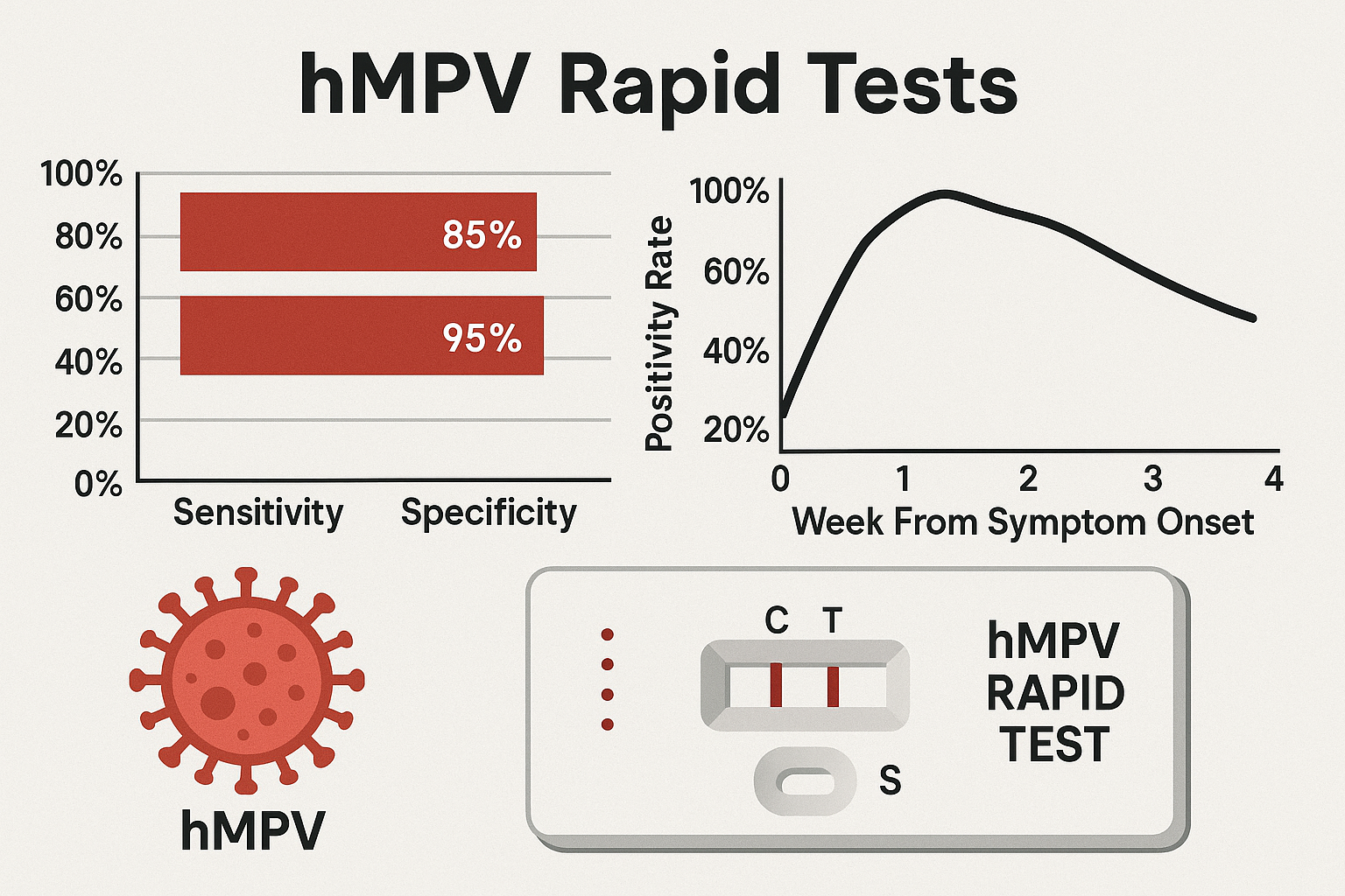
Multiple studies, such as those archived in the Journal of Clinical Microbiology (ASM), have compared rapid antigen assays with RT-PCR and immunofluorescence. The hMPV Rapid Test exhibits:

  • Sensitivity between 85–95%

  • Specificity above 98%

  • Cross-reactivity testing against RSV, Adenovirus, and Influenza with no false positives

The Clinical Laboratory Improvement Amendments (CLIA) framework requires ongoing internal quality control, and FDA Emergency Use Authorizations (EUA) guidelines specify performance metrics for respiratory antigen tests.

Clinical and Research Applications

Rapid identification of hMPV is valuable in multiple diagnostic contexts:

  • Distinguishing hMPV from bacterial pneumonia to reduce unnecessary antibiotics

  • Screening hospitalized patients during respiratory outbreaks

  • Surveillance studies in pediatric and geriatric populations

  • Use in vaccine development and viral pathogenesis research

According to data in the National Library of Medicine, hMPV is frequently detected alongside RSV, rhinoviruses, or SARS-CoV-2, making multiplex testing panels a preferred approach in advanced laboratories. Institutions such as Stanford Medicine and Johns Hopkins University have published studies demonstrating the role of combined antigen panels in accurate respiratory virus monitoring.

Advantages of Rapid Antigen Testing

The U.S. Department of Health and Human Services (HHS) outlines the major benefits of point-of-care testing for respiratory viruses:

  • Rapid isolation and containment measures

  • Cost-effective outbreak control

  • Support for epidemiological data collection

  • Simplified logistics in low-resource settings

For pediatric health, the American Academy of Pediatrics (AAP) supports antigen testing as a front-line screening tool for respiratory infections, emphasizing its importance in daycare and hospital environments.

Limitations and Confirmatory Testing

While highly efficient, antigen-based assays are inherently less sensitive than nucleic acid amplification tests (NAATs). Therefore, negative hMPV rapid results in symptomatic patients should be confirmed by RT-PCR, following recommendations by the European Centre for Disease Prevention and Control (ECDC).
External quality assessment schemes can be conducted under supervision of the CDC Laboratory Quality Program and CLSI Standards.

Integration in Multiplex Respiratory Panels

Modern diagnostics integrate hMPV antigen detection into multiplex immunochromatographic panels, allowing simultaneous identification of Influenza A/B, RSV, and hMPV.
These platforms, validated under FDA EUA listings, provide broad coverage and real-time surveillance capability.

Research at MIT Department of Biological Engineering and NIST explores nanostructured membranes and signal-amplified assays that could further enhance analytical sensitivity of hMPV antigen detection.

Test Procedure (General Workflow)

  1. Bring the test device and sample to room temperature.

  2. Add extraction buffer to the tube and insert the swab sample.

  3. Mix thoroughly to release viral antigens.

  4. Add 3–4 drops of extracted solution into the sample well of the test cassette.

  5. Read results visually after 10–15 minutes.

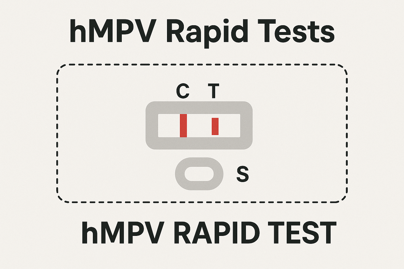
The appearance of two colored lines indicates a positive result, while one control line denotes a negative. Absence of any line signifies an invalid test that should be repeated.

Detailed operating instructions are outlined by the CDC Laboratory Training Network and FDA Device Labeling Guidance.

Storage, Stability, and Shelf Life

According to the WHO Laboratory Quality Management System, rapid test kits should be stored at 2–30 °C in a dry environment. Avoid freezing, as low temperatures can denature antibodies.
Once opened, test cassettes must be used immediately. The shelf life, typically 18–24 months, should be verified through the lot-specific Certificate of Analysis (CoA) provided by the manufacturer.

Data Interpretation and Reporting

For accurate data reporting, laboratories can integrate hMPV Rapid Test results into digital systems following guidelines from the National Institute for Health Data Science (NIHDS).
Standardized coding systems such as LOINC and SNOMED CT facilitate interoperability between laboratory information management systems (LIMS) and clinical databases.

Future Innovations in hMPV Diagnostics

Emerging diagnostic trends include:

  • Microfluidic assay miniaturization

  • Quantum-dot-based fluorescent detection

  • AI-driven image analysis for visual line quantification

  • Integration with smartphone-based readers for decentralized diagnostics

Institutions such as the National Science Foundation (NSF) and NIH Rapid Acceleration of Diagnostics (RADx) Initiative are supporting projects aimed at revolutionizing rapid respiratory testing.

Regulatory Framework and Compliance

While non-medical blogs should avoid clinical guidance, it is important to note that hMPV rapid assays adhere to IVD regulatory frameworks defined by the FDA, ISO 13485, and CE marking in Europe.
These regulations ensure consistency, reproducibility, and safety in diagnostic manufacturing and distribution.

Educational and Research Resources

Researchers interested in hMPV pathogenesis and diagnostic assay development can consult:

These resources support academic understanding and facilitate transparent communication between research and diagnostic sectors.

Conclusion

The hMPV Rapid Test represents a critical advancement in modern diagnostic science — delivering speed, accuracy, and accessibility for viral detection. Its design simplifies laboratory workflows, supports global surveillance efforts, and bridges the gap between traditional molecular diagnostics and real-time point-of-care analysis.

As technology continues to evolve through the collaboration of institutions like the NIH, CDC, and NIST, the future of hMPV detection lies in smarter, more sensitive, and digitally connected platforms. These innovations ensure that laboratories, biotechnologists, and research professionals maintain the highest standards in viral diagnostics.

Explore the technical foundation and diagnostic importance of hMPV Rapid Tests — advanced lateral-flow assays for human metapneumovirus detection. Learn about test principles, accuracy, and future innovations from verified .edu and .gov resources.