Avian Infectious Bursal Disease: Molecular Characteristics, Avian Immunology Research, and Laboratory Study Approaches

Introduction

Avian Infectious Bursal Disease (IBD), widely known in poultry science as Gumboro disease, represents one of the most extensively studied viral conditions affecting chickens and other avian species. In research settings, IBD has become an important experimental model for understanding avian immunology, viral pathogenesis, and lymphoid tissue biology.

The virus responsible for this condition, Infectious Bursal Disease Virus (IBDV), selectively targets the bursa of Fabricius, a specialized immune organ unique to birds. Because the bursa plays a central role in B-lymphocyte maturation, the virus provides a valuable system for exploring the molecular mechanisms that regulate immune development.

Researchers studying avian virology, veterinary immunology, molecular biology, and poultry science frequently analyze IBDV in order to better understand:

  • RNA virus evolution

  • host–pathogen interactions

  • immune cell differentiation

  • viral replication mechanisms

Educational resources explaining the biology of poultry viruses and avian immune systems can be explored through the United States Department of Agriculture (USDA)
https://www.usda.gov

Additional academic materials on poultry diseases and avian biology are available from the University of Georgia Poultry Science Department
https://poultry.caes.uga.edu/

Veterinary virology training and research programs can also be found at the Cornell University College of Veterinary Medicine
https://www.vet.cornell.edu/

Historical Background of Infectious Bursal Disease Research

The disease now referred to as Infectious Bursal Disease was first described in the early 1960s near the town of Gumboro, Delaware, which led to the commonly used name Gumboro disease. Since its initial identification, the virus has attracted major scientific interest because of its unique effect on avian lymphoid tissues.

Over time, research laboratories around the world have investigated the molecular biology of IBDV, leading to significant discoveries regarding viral genome organization, viral capsid structure, and immune system interactions.

Historical and scientific documentation on avian viral diseases can be explored through the National Library of Medicine
https://www.nlm.nih.gov/

Comprehensive genomic research databases are maintained by the National Center for Biotechnology Information (NCBI)
https://www.ncbi.nlm.nih.gov/

Educational materials on RNA viruses and viral genome structure are also available from the National Institutes of Health (NIH)
https://www.nih.gov/

Taxonomy and Classification of Infectious Bursal Disease Virus

The causative agent of Avian Infectious Bursal Disease is Infectious Bursal Disease Virus (IBDV), a member of the Birnaviridae family.

Viruses belonging to this family share several defining characteristics:

  • Non-enveloped viral particles

  • Double-stranded RNA genomes

  • Segmented genomic structure

  • Icosahedral capsid symmetry

IBDV is classified within the genus Avibirnavirus, which includes viruses that specifically infect avian hosts.

Taxonomic classification of viruses is maintained by international virology databases such as those described by the National Institutes of Health
https://www.nih.gov/

Further educational resources on virus classification are provided by MIT Biology
https://biology.mit.edu/

Researchers studying viral genome structure can also access molecular virology resources from Stanford University School of Medicine
https://med.stanford.edu/

Molecular Structure of IBDV

The genome of Infectious Bursal Disease Virus is divided into two double-stranded RNA segments, referred to as Segment A and Segment B.

Segment A

Segment A encodes several viral proteins including:

  • VP2 – major capsid protein involved in antigenicity

  • VP3 – structural protein interacting with viral RNA

  • VP4 – viral protease responsible for polyprotein processing

  • VP5 – non-structural protein associated with virus release

Segment B

Segment B encodes VP1, the RNA-dependent RNA polymerase, which is essential for viral genome replication.

Detailed genomic information about viral RNA replication can be explored through the National Human Genome Research Institute
https://www.genome.gov/

Educational virology programs examining viral genome replication are conducted by the University of California Davis School of Veterinary Medicine
https://www.vetmed.ucdavis.edu/

The Bursa of Fabricius and Avian Immune System Development

A defining feature of IBDV research is the virus’s interaction with the bursa of Fabricius, an immune organ located near the cloaca of birds.

The bursa is responsible for B-cell maturation, a process essential for antibody production in the avian immune system.

Because of this biological role, the bursa has become an important focus in research areas such as:

  • immune organ development

  • lymphocyte differentiation

  • adaptive immune system evolution

Educational resources explaining immune system development are provided by Harvard University’s Department of Molecular and Cellular Biology
https://mcb.harvard.edu/

Further academic material on avian immunology is available through North Carolina State University Poultry Science Program
https://poultry.ces.ncsu.edu/

Additional immunology research programs can be explored at Yale School of Medicine
https://medicine.yale.edu/
AffiVET® Avian Infectious Bursal Disease (IBD) Antibody Elisa Test Kit

Viral Replication and Host–Virus Interactions

The replication cycle of Infectious Bursal Disease Virus has been extensively studied in molecular virology laboratories.

The viral replication process generally involves:

  1. Attachment of the virus to host cells

  2. Entry into lymphoid cells within the bursa

  3. Release of viral RNA genome

  4. Replication by RNA-dependent RNA polymerase

  5. Assembly of new viral particles

  6. Release from infected cells

These processes allow researchers to explore fundamental biological questions about RNA virus replication and host cell responses.

Educational information about viral replication strategies can be accessed through Johns Hopkins University School of Medicine
https://www.hopkinsmedicine.org/

Further molecular virology resources are available from University of Michigan Medical School
https://medicine.umich.edu/

Global Research on Avian Viral Pathogens

Because poultry production represents a major agricultural sector worldwide, avian viral diseases are widely investigated by research institutions and agricultural laboratories.

International organizations supporting poultry health research include the Food and Agriculture Organization of the United Nations (FAO)
https://www.fao.org/

Global animal health programs are coordinated by the World Organisation for Animal Health (WOAH)
https://www.woah.org/

These organizations collaborate with universities and veterinary institutes to advance scientific understanding of poultry pathogens.

Additional research information about agricultural animal health can be found through the United States Department of Agriculture Animal and Plant Health Inspection Service
https://www.aphis.usda.gov/

Laboratory Techniques Used in IBD Research

Research laboratories studying Avian Infectious Bursal Disease Virus use multiple analytical methods to investigate viral biology and immune responses.

ELISA-Based Research Methods

Enzyme-Linked Immunosorbent Assay (ELISA) techniques are widely used in research laboratories to study viral proteins or immune responses associated with IBDV.

Educational descriptions of immunoassay technologies are available from the National Institute of Biomedical Imaging and Bioengineering
https://www.nibib.nih.gov/

Additional laboratory immunology resources are available from University of Washington Department of Immunology
https://immunology.washington.edu/

PCR and Molecular Detection

Polymerase Chain Reaction (PCR) techniques are commonly used in molecular virology research to analyze viral RNA and characterize viral strains.

Educational PCR resources are provided by the National Science Foundation
https://www.nsf.gov/

Further molecular genetics training materials are available through Stanford University Genetics Department
https://med.stanford.edu/genetics.html

Histopathological Analysis

Histological examination of bursal tissue allows researchers to study structural changes in lymphoid organs.

Microscopy and tissue analysis training programs are available at the University of Pennsylvania School of Veterinary Medicine
https://www.vet.upenn.edu/

Additional pathology education resources are provided by Iowa State University Veterinary Diagnostic Laboratory
https://www.vetmed.iastate.edu/

Genomic and Bioinformatics Research

Advances in genomic sequencing and computational biology have greatly expanded the study of IBDV genetic diversity.

Researchers now analyze viral genomes to investigate:

  • genetic variation among viral strains

  • mutation patterns in viral capsid proteins

  • evolutionary relationships among viruses

Bioinformatics databases used in viral genomics research are maintained by the European Bioinformatics Institute
https://www.ebi.ac.uk/

Additional genomic research tools are available from the National Institutes of Health Genome Data Science programs
https://datascience.nih.gov/

Research Applications in Poultry Science

Research tools designed for studying Avian Infectious Bursal Disease support a wide variety of scientific investigations.

These investigations often focus on:

  • immune system development in birds

  • viral protein structure and antigenicity

  • RNA virus replication mechanisms

  • host immune signaling pathways

Academic programs studying poultry biology and veterinary science are conducted by institutions such as Texas A&M University College of Veterinary Medicine
https://vetmed.tamu.edu/

Further poultry research programs are available at Auburn University Poultry Science Department
https://poultry-science.auburn.edu/

Importance of High-Quality Laboratory Research Reagents

Reliable laboratory reagents play an essential role in ensuring consistent and reproducible scientific experiments.

Research products designed for the investigation of Avian Infectious Bursal Disease support laboratory workflows involving:

  • viral antigen detection

  • molecular biology experiments

  • immunological assays

  • viral genome analysis

Laboratory biosafety and research standards are described by the NIH Office of Science Policy
https://osp.od.nih.gov/

Guidelines for laboratory safety practices are also provided by the Centers for Disease Control and Prevention Biosafety Program
https://www.cdc.gov/labs/BMBL.html

Conclusion

Avian Infectious Bursal Disease remains one of the most important subjects of investigation in poultry science, veterinary virology, and avian immunology research. The virus’s selective interaction with the bursa of Fabricius and developing B-cells makes it a valuable experimental model for studying immune system biology.

Through the use of modern molecular biology tools, immunological assays, and genomic sequencing technologies, scientists continue to expand our understanding of IBDV structure, replication mechanisms, and host immune responses.

Research tools designed for studying Avian Infectious Bursal Disease support universities, veterinary research institutes, and agricultural laboratories worldwide as they explore the complex biology of avian viral pathogens.
Infectious Bursal Disease Virus research
IBDV molecular biology
avian immunology research
poultry virology studies
bursa of Fabricius research
avian viral pathogen research
poultry virus laboratory studies
IBDV genomic analysis
avian immunology laboratory research

50× ROX Reference Dye: Technical Overview, Use & Importance in qPCR

Introduction to 50× ROX Reference Dye

50× ROX Reference Dye, also known as carboxy-X-rhodamine (ROX), is one of the most commonly used reference dyes in quantitative PCR (qPCR). It plays a pivotal role in improving the accuracy and reliability of qPCR results by normalizing fluorescence signals during the amplification process. This normalization helps correct for non-PCR-related variations, such as optical path length differences, slight variations in volume, or even the inherent fluorescence variability of the PCR system.

ROX is typically provided in a 50× concentration (approximately 25 µM), which must be diluted to a final working concentration depending on the specific qPCR platform being used. Despite its lack of direct involvement in the amplification reaction, ROX serves as an invaluable passive reference to maintain consistency and correct for various technical errors during data acquisition.

This article will provide an in-depth overview of 50× ROX Reference Dye, its critical role in qPCR, best practices for its use, troubleshooting tips, and real-world applications in molecular biology research, diagnostics, and biotechnology.

For a detailed guide on quantitative PCR, visit NIH’s PCR section.

AffiGEN® 50x ROX Reference Dye

What is ROX Reference Dye and Why is it Used in qPCR?

ROX Reference Dye is a passive reference dye, meaning it does not participate in the amplification process. It does not bind to DNA or interfere with the polymerase, making it a highly stable and reliable signal used for normalization. As an inert substance, ROX maintains a consistent fluorescence signal throughout the entire amplification cycle, irrespective of the amount of target DNA present in the reaction.

The 50× stock solution of ROX must be diluted to a working concentration before adding it to the PCR mix. The final concentration depends on the specific type of qPCR machine and its sensitivity. For instance, high-ROX and low-ROX concentrations are often used depending on the machine’s optical system and the user’s specific experimental needs.

How ROX Reference Dye Works

In qPCR, the fluorescence emitted from specific probes or DNA-binding dyes is measured as a function of the amount of amplified product present in each cycle. However, various factors, such as:

  • Differences in well-to-well fluorescence

  • Instrument variation

  • Changes in reaction volume during thermal cycling

can introduce inaccuracies into the measurement. ROX corrects for these variations by serving as a constant fluorescence signal throughout the experiment. When fluorescence readings are taken, the system normalizes the signal of the target reporter dye (such as SYBR Green or TaqMan probes) to the stable signal of ROX. This normalization ensures that the fluorescence measured reflects only the amplification of the target DNA, not variations due to other factors.

The formula for normalization is:
Rn = (Fluorescence of reporter dye) / (Fluorescence of ROX)

This ratio, called ΔRn, is used to generate the amplification curve for real-time PCR experiments.

Benefits of Using 50× ROX Reference Dye

  1. Data Normalization: ROX helps adjust for optical and physical variations between wells in a multi-well plate, ensuring that data collected across all wells is comparable and accurate.

  2. Improved Reproducibility: By normalizing fluorescence, ROX ensures that results from different PCR plates or runs can be compared without needing to account for minor system-specific differences in fluorescence intensity.

  3. Enhanced Multiplexing: ROX enables successful multiplex assays by ensuring that the signal from each reporter dye used in the multiplex reaction is normalized against a consistent reference signal, avoiding interference between different fluorescence channels.

  4. Reduction of Technical Variation: External factors such as evaporation, condensation, and pipetting errors can cause variability in qPCR. ROX accounts for these discrepancies, ensuring the reproducibility of results.

  5. Time and Cost Efficiency: By reducing the need for multiple replicates or technical corrections, the use of ROX can make experiments more efficient, both in terms of time and cost.

For an example of how ROX improves multiplex assay accuracy, consult Thermo Fisher’s guide on multiplex PCR.

Determining the Correct ROX Concentration: High ROX vs. Low ROX

50× ROX can come in high ROX and low ROX formulations, which differ based on the required final concentration for the instrument being used. The concentration of ROX needed depends largely on the optical system of the qPCR platform.

Low ROX Concentration (50 nM)

  • Typically used with machines that have low fluorescence sensitivity or advanced optical designs that minimize well-to-well variation.

  • Instruments such as the Bio-Rad CFX96 and Thermo Fisher QuantStudio series often use low ROX.

High ROX Concentration (500 nM)

  • Suitable for older machines or those with high sensitivity, where the instrument requires a higher signal to normalize against.

  • Instruments like the Applied Biosystems 7500 Fast require high ROX concentrations for optimal performance.

Make sure to verify the required concentration for your machine by consulting the manufacturer’s documentation. For a detailed comparison of instruments and their ROX requirements, visit Applied Biosystems’ PCR platform guide.

Best Practices for Using 50× ROX in qPCR

  1. Proper Dilution: Always dilute the 50× ROX stock solution to the appropriate final concentration based on your instrument’s needs. Overuse or underuse of ROX can lead to inaccurate normalization and poor data quality.

  2. Mixing Thoroughly: ROX should be thoroughly mixed into the PCR master mix. This ensures that it is evenly distributed and maintains a consistent signal across all wells. Vortexing and quick pipetting can ensure homogeneous mixing.

  3. Add ROX After Preparing Master Mix: Add ROX to the reaction mix after preparing the master mix but before aliquoting it into individual PCR tubes or plates. This ensures even distribution and avoids any concentration discrepancies.

  4. Protection from Light: ROX is a fluorescent dye, so it should be stored in the dark and handled in low-light conditions to avoid photo-bleaching. Always store it at –30 °C to –10 °C to maintain stability.

  5. Reagent Compatibility: Ensure that the other reagents in your PCR master mix do not interact negatively with ROX. Some additives, buffers, or primers may affect the fluorescence of ROX.

For a detailed step-by-step guide, visit Bio-Rad’s best practices.

Advanced Applications of 50× ROX Reference Dye

Multiplex qPCR Assays

Multiplex qPCR allows the simultaneous amplification and detection of multiple targets in the same PCR reaction. ROX plays an essential role in multiplexing by normalizing the signals from different reporter dyes used in the assay. For instance, researchers can amplify multiple targets with distinct fluorescent probes (e.g., FAM, VIC, or Cy5), while using ROX as a consistent internal reference to ensure accurate quantification of each target without spectral overlap.

Pathogen Detection

In diagnostics, especially in pathogen detection, accurate quantification is critical. ROX is commonly used in assays designed to detect viral or bacterial DNA or RNA, ensuring the fluorescence from target-specific probes is accurately measured, irrespective of minor technical variations that may arise in the laboratory setup.

For example, assays like the TaqMan COVID-19 detection kit or assays for Enterovirus detection often employ ROX for high-precision results in clinical settings.

For more about pathogen detection techniques and their challenges, refer to CDC’s guidance on diagnostic PCR assays.

Gene Expression Analysis

In gene expression studies, ROX allows for the precise quantification of target genes by correcting for inconsistencies that could affect the interpretation of results. This is especially important when comparing the relative expression of genes across different samples, where accurate baseline normalization is essential.

For guidance on setting up gene expression studies with proper normalization, see NIH’s gene expression resource.

Troubleshooting Common Issues with ROX

While ROX Reference Dye is generally straightforward to use, researchers may encounter some common issues. Below are troubleshooting tips:

  1. Inconsistent Data Normalization: If you notice that data normalization seems unreliable or the baseline correction is poor, check if the correct ROX concentration has been used. Overdilution or underdilution can lead to improper normalization. Always double-check your instrument’s requirements.

  2. Fluorescence Saturation: Using too high of a concentration of ROX (especially in high-ROX instruments) can cause fluorescence saturation, leading to flat or unresponsive amplification curves. Ensure you’re following manufacturer guidelines on optimal concentrations.

  3. Fluorescence Background Noise: If background noise appears elevated, it could be a sign of interference from ROX or other reagents. Try adjusting the concentration or testing with a different passive reference dye, if necessary.

  4. Data Anomalies Due to Evaporation: In experiments with high sensitivity, evaporation in wells may still cause discrepancies, even with ROX normalization. To address this, ensure that plates are sealed properly and use the correct volume of reagents.

For more troubleshooting tips, check out Thermo Fisher’s PCR troubleshooting guide.

Conclusion: The Essential Role of 50× ROX Reference Dye in Real-Time PCR

50× ROX Reference Dye is an indispensable tool for ensuring accurate, reproducible, and reliable results in real-time PCR. By normalizing fluorescence signals, ROX accounts for variations that could otherwise introduce errors in the quantification of nucleic acids. Whether in gene expression analysis, pathogen detection, or multiplex assays, ROX enables high-quality, reliable data from qPCR experiments.

Adhering to best practices for the proper use of ROX — such as correct dilution, mixing, and ensuring compatibility with the PCR platform — is key to achieving optimal results. While most users encounter minimal issues with ROX, understanding its role and troubleshooting any potential issues ensures researchers can confidently rely on this passive reference dye for their most important experimental results.

For further reading and detailed technical insights, please refer to authoritative resources like NIH’s PCR guide, Thermo Fisher’s resource center, and Bio-Rad’s PCR manual.

Enterovirus A71 (EV71) RNA PCR Quantitative Positive Control — Complete Scientific and Technical Review

Introduction

Enterovirus A71 (EV71) is a non-enveloped, positive-sense single-stranded RNA virus belonging to the Picornaviridae family.
It is one of the primary etiological agents of Hand, Foot and Mouth Disease (HFMD) and is responsible for severe neurological complications, such as:

  • Brainstem encephalitis

  • Acute flaccid paralysis

  • Neurogenic pulmonary edema

  • Meningitis in children

Accurate detection of EV71 RNA is essential for clinical diagnostics, public-health surveillance, and laboratory research.

The EV71 RNA PCR Quantitative Positive Control is a high-precision reference material used to:

  • Validate qPCR assays

  • Confirm detection sensitivity

  • Assess analytical accuracy

  • Support assay calibration and quantification

  • Verify instrument performance (CT linearity, fluorescence response)

  • Ensure reliable diagnostic workflows for EV71 molecular testing

This article provides a full, long-form scientific review including molecular biology, PCR assay design, positive-control engineering, biosafety, quantification methods, and QC integration.

AffiCHECK® Enterovirus A71 (EV71) RNA PCR Quantitative Positive Control

Molecular Biology of Enterovirus A71 (EV71)

 Genome Structure

EV71 contains a ~7.4 kb positive-sense ssRNA genome with:

  • 5’ UTR region (highly structured, ideal for PCR targeting)

  • Single ORF encoding polyprotein (P1, P2, P3)

  • 3’ UTR + poly(A) tail

The polyprotein is processed into:

  • Structural proteins: VP1, VP2, VP3, VP4

  • Nonstructural proteins: 2A, 2B, 2C, 3A, 3B, 3Cpro, 3Dpol

The VP1 region is highly relevant for typing and molecular detection.

Virological Significance

EV71 is associated with periodic epidemics, especially in:

  • Southeast Asia

  • Western Pacific Region

  • Emerging clusters worldwide

Because of its neurotropic profile, detecting the virus early using qRT-PCR is crucial.

Principles of EV71 RNA Detection by qRT-PCR

 Why qRT-PCR?

Real-time reverse transcription PCR (qRT-PCR) is considered the gold standard for viral RNA detection because it offers:

  • High sensitivity

  • High specificity

  • Low limit of detection (LOD)

  • Rapid turnaround time

  • Compatibility with clinical and environmental samples

 Target Regions Used in PCR Assays

Common qPCR targets for EV71 include:

  • 5’ UTR (broad enterovirus detection)

  • VP1 (type-specific, high accuracy)

  • 3Dpol (polymerase gene, conserved)

An EV71 Positive Control typically mirrors the chosen assay target.

What Is an EV71 RNA PCR Quantitative Positive Control?

A Quantitative Positive Control is a carefully engineered RNA standard containing the EV71 target sequences used in qRT-PCR.

It allows laboratories to:

  • Validate assay sensitivity and accuracy

  • Ensure reagent and instrument performance

  • Establish quantification curves

  • Detect degradation or inhibition in extraction/PCR workflows

  • Standardize inter-laboratory comparison

 Forms of EV71 Positive Controls

  • Synthetic RNA transcripts

  • In vitro transcribed viral RNA fragments

  • Armored RNA (RNase-resistant encapsulated RNA)

  • Plasmid or synthetic gene standards (used after reverse transcription)

  • Whole inactivated virus (for extraction controls — BSL considerations apply)

Most commercial positive controls are non-infectious synthetic RNA, ensuring biosafety.

Design and Engineering of EV71 Positive Control RNA

https://media.springernature.com/lw685/springer-static/image/art%3A10.1038%2Fs41598-021-81760-0/MediaObjects/41598_2021_81760_Fig1_HTML.png

 Sequence Selection

The control includes:

  • Highly conserved EV71 sequences

  • PCR primer & probe binding sites

  • Calibration sequences for quantification

 In Vitro Transcription

Process typically uses:

  • Linearized plasmid with EV71 insert

  • T7 or SP6 RNA polymerase

  • NTP substrate mixture

  • DNase step to remove template DNA

 Quantification

Controls are provided with precise viral copy numbers, e.g.:

  • 10⁶ copies/µL

  • 10⁵ copies/µL

  • 10⁴ copies/µL

  • 10³ copies/µL

This allows creation of standard curves to validate CT accuracy and dynamic range.

 Stability Enhancements

High-end positive controls may include:

  • RNase inhibitors

  • Encapsulation (armored RNA)

  • Lyophilization for room-temperature shipping

  • Cryoprotectants for long-term stability

Applications of EV71 RNA Quantitative Positive Controls

 Clinical qRT-PCR Assays

Used in routine clinical detection for:

  • Throat swabs

  • Vesicle fluid

  • CSF samples

  • Stool samples

Ensures:

  • Sensitivity validation (LOD verification)

  • Target specificity confirmation

  • Accuracy of CT values

 Quality Assurance & QC Programs

Ideal for:

  • Lot-to-lot comparison

  • Internal QC

  • External quality assessment (EQA)

  • Proficiency testing

 Research Applications

  • Viral load quantification

  • Variant research

  • Primer/probe evaluation

  • Cross-reactivity assessment

  • Extraction efficiency testing

 Instrument Calibration

Used to validate:

  • CT linearity

  • Fluorescence detection

  • Thermal cycling accuracy

  • RT efficiency

Technical Advantages of High-Quality EV71 Controls

  • Non-infectious

  • Highly stable RNA

  • Exact viral copy quantification

  • Compatible with TaqMan, SYBR Green, multiplex PCR

  • Suitable for digital PCR (dPCR)

  • Guaranteed reproducibility across experiments

  • Works with all major platforms:

    • ABI

    • Bio-Rad

    • Roche LightCycler

    • Qiagen Rotor-Gene

    • QuantStudio Series

    • OpenArray

Biosafety and Handling Considerations

Although EV71 synthetic positive controls are non-infectious, good laboratory practices apply:

  • Use RNase-free consumables

  • Wear gloves & lab coat

  • Avoid freeze–thaw cycles

  • Store at −80°C (or −20°C for lyophilized forms)

  • Aliquot RNA immediately after reconstitution

  • Protect from light and RNase contamination

Preparation of Standard Curves Using EV71 Positive Controls

A proper standard curve includes:

  • Serial dilutions (10⁶ → 10¹ copies)

  • Triplicate CT readings

  • R² ≥ 0.98

  • PCR efficiency 90–105%

This validates:

  • Assay sensitivity

  • Reproducibility

  • Limit of detection

  • Quantification accuracy

https://www.researchgate.net/publication/288872451/figure/fig1/AS%3A614263852892160%401523463323100/Structure-and-genome-of-Enterovirus-71-The-capsid-consists-of-60-protomers-each.png

Troubleshooting Using EV71 Positive Controls

 High CT Values

  • RNA degradation

  • Incorrect dilution

  • Poor RT efficiency

 No Amplification

  • Primer/probe mismatch

  • PCR master mix failure

  • Thermocycler issues

 Inconsistent CT Values

  • Pipetting error

  • RNA adsorption on plastic

  • Inhibition in reaction mixture

A quantitative control is essential for diagnosing these issues.

Conclusion

The Enterovirus A71 (EV71) RNA PCR Quantitative Positive Control is an indispensable component of modern molecular diagnostics and virology research.

It ensures that qRT-PCR assays are:

  • Accurate

  • Sensitive

  • Reproducible

  • Quantitative

  • Globally standardized

By providing a precisely quantified, stable, and safe RNA standard, EV71 positive controls empower laboratories to detect enteroviral infections rapidly and reliably — supporting clinical diagnostics, surveillance programs, vaccine development, extraction QC, and instrument calibration.

Throughout the article, the strongest indexing keywords for Google include:

  • Enterovirus A71 EV71 RNA Positive Control

  • EV71 qRT-PCR quantitative standard

  • Hand Foot and Mouth Disease molecular detection

  • Enterovirus RT-PCR calibration control

  • EV71 VP1 RNA standard curve

  • Armored RNA EV71 positive control

  • EV71 PCR assay validation

  • Enterovirus A71 reference material

Molgramostim (Recombinant Human GM-CSF): An In-Depth Scientific Review of Structure, Mechanism, Immunological Function, Bioprocessing, and Clinical Relevance

INTRODUCTION

Molgramostim is the E. coli-derived recombinant form of human GM-CSF (Granulocyte-Macrophage Colony-Stimulating Factor), a key hematopoietic cytokine that regulates innate immunity, myelopoiesis, dendritic cell differentiation, and inflammatory signaling.

GM-CSF plays an essential role in the:

  • Survival, proliferation, and maturation of granulocyte and macrophage precursors

  • Activation of mature neutrophils, monocytes, and macrophages

  • Maturation of dendritic cells (DCs)

  • Orchestration of adaptive immune priming

  • Clearance of pulmonary surfactant (critical in lung immunity)

As a non-glycosylated recombinant cytokine, Molgramostim matches the amino acid sequence of natural human GM-CSF, maintaining full bioactivity while offering high purity, stability, and batch consistency — characteristics crucial for research applications in immunology, oncology, vaccine development, and hematology.

Because GM-CSF sits at the crossroads of immunoregulation, hematopoiesis, cancer immunotherapy, and pulmonary disease biology, Molgramostim is considered one of the most important experimental cytokines of the past three decades.

AffiREC® Molgramostim: Recombinant Human GM-CSF

GM-CSF: Molecular Architecture and Biological Identity

 Structural Organization

GM-CSF is a four-helix bundle cytokine, approximately 14.5 kDa, stabilized by:

  • Two conserved disulfide bridges (Cys54-Cys96 and Cys88-Cys121)

  • Hydrophobic packing within the helical bundles

  • Receptor-binding loops on helices α1 and α4

The recombinant version, Molgramostim, is non-glycosylated because E. coli does not perform eukaryotic glycosylation.
Despite this, the absence of glycan chains:

  • Does not compromise receptor binding

  • Causes a slightly shorter in vivo half-life

  • Provides excellent reproducibility for in vitro studies

 GM-CSF Receptor System

GM-CSF signals through a heterodimeric receptor complex consisting of:

  • GM-CSF-Rα (CD116) → high specificity

  • βc (CD131) → shared with IL-3 and IL-5

Receptor engagement triggers structural rearrangements that initiate downstream signaling cascades.

GM-CSF Signal Transduction Pathways

Upon binding, Molgramostim activates multiple intracellular pathways:

JAK2-STAT5 Axis (Primary Pathway)

  • Receptor dimerization activates JAK2

  • JAK2 phosphorylates STAT5

  • STAT5 translocates to the nucleus

  • Induces genes supporting:

    • Proliferation

    • Anti-apoptosis

    • Myeloid cell differentiation

MAPK Pathway (ERK1/2)

Leads to:

  • Cellular survival

  • Enhanced cytokine secretion

  • Proliferation of progenitors

PI3K-AKT Pathway

Controls:

  • Cell survival

  • Metabolic adaptation

  • Anti-apoptotic signaling

NF-κB Activation

Critical for:

  • Macrophage inflammatory responses

  • Antigen presentation

  • Cytokine amplification

These pathways collectively enable GM-CSF to shape the innate and adaptive immune landscape.

https://i.shgcdn.com/782e2c99-efc6-4a47-bb52-4e731efdae83/-/format/auto/-/preview/3000x3000/-/quality/lighter/

Functional Biology of Molgramostim

Myelopoiesis and Hematopoietic Recovery

Molgramostim increases:

  • Neutrophil production

  • Macrophage maturation

  • Eosinophil and basophil activation

  • Expansion of bone marrow CFU-G, CFU-M, and CFU-GM colonies

This makes it vital for studying:

  • Chemotherapy-induced myelosuppression

  • Bone marrow transplantation

  • Hematopoietic stem cell mobilization

Macrophage Activation and Immune Amplification

Molgramostim enhances:

  • Phagocytosis

  • ROS/RNS production

  • Cytokine secretion (TNF-α, IL-1β, IL-6, IL-23)

  • Microbicidal capacity

  • Antigen presentation

These characteristics position GM-CSF as a major “immune amplifier.”

Dendritic Cell Differentiation

Molgramostim, together with IL-4, is the gold standard for generating:

  • Monocyte-Derived Dendritic Cells (MoDC)

  • Mature antigen-presenting dendritic cells

  • DC vaccines used in cancer research

Lung Immunity and Surfactant Homeostasis

GM-CSF is essential for alveolar macrophage development and:

  • Surfactant catabolism

  • Pathogen clearance

  • Lung homeostasis

A deficiency in GM-CSF signaling is the root cause of Pulmonary Alveolar Proteinosis (PAP).

Production of Molgramostim (Recombinant Human GM-CSF)

 Gene Design & Cloning

The human CSF2 gene is:

  • Codon-optimized for E. coli

  • Inserted into high-copy expression vectors

  • Transformed into production strains like BL21(DE3)

 Expression in E. coli

GM-CSF often aggregates into inclusion bodies, offering advantages:

  • Higher purity

  • Ease of isolation

  • Protection from proteolysis

 Protein Refolding and Purification

To recover active cytokine:

  1. Inclusion bodies are solubilized with chaotropic agents

  2. Refolding occurs via step-wise dialysis

  3. Purification uses:

    • HPLC

    • Ion-exchange chromatography

    • Size-exclusion chromatography

 Quality Control and Analytics

QC testing includes:

  • SDS-PAGE purity (>95%)

  • Endotoxin testing

  • Mass spectrometry

  • Circular dichroism folding analysis

  • Bioassays for GM-CSF activity

Molgramostim vs Sargramostim (Key Scientific Comparison)

Feature Molgramostim (E. coli) Sargramostim (Yeast)
Glycosylation None Hyperglycosylated
Size ~14.5 kDa ~19 kDa
Bioactivity Highly potent in vitro Slightly altered receptor kinetics
Immunogenicity Low Possible yeast glycan immunogenicity
Research preference Excellent for controlled assays Used clinically

Molgramostim is preferred in research environments due to its:

  • uniform structure

  • high batch reproducibility

  • reliable receptor interaction

GM-CSF receptor and signaling. | Download Scientific Diagram

Applications in Biomedical & Clinical Research

 Oncology and Immunotherapy

Molgramostim is central in studies of:

  • Dendritic cell vaccines

  • Tumor-associated macrophage (TAM) reprogramming

  • Checkpoint inhibitor combination therapy

  • Oncolytic viruses expressing GM-CSF (e.g., modified HSV-1)

  • T-cell expansion and priming

 Vaccine Science

GM-CSF acts as a biological adjuvant, enhancing:

  • APC recruitment

  • Th1/Th17 polarization

  • CD8+ T-cell priming

  • Antibody responses

Used in:

  • Cancer neoantigen vaccines

  • Viral vaccines

  • DNA/RNA vaccine platforms

Hematology and Bone Marrow Biology

Molgramostim is widely used for:

  • Stem cell mobilization studies

  • Hematopoietic niche modeling

  • Bone marrow organoid development

 Pulmonary Alveolar Proteinosis (PAP)

GM-CSF corrects the macrophage defect responsible for surfactant accumulation.

Infectious Disease Immunology

GM-CSF enhances macrophage defenses against:

  • Bacteria

  • Fungi

  • Viruses

  • Intracellular pathogens

Formulation, Storage, and Handling

Molgramostim is supplied as lyophilized powder:

  • Store at −20°C to −80°C

  • Reconstitute with sterile water + carrier protein (BSA/HSA)

  • Avoid freeze–thaw cycles

  • Typical working concentration: 1–50 ng/mL

Advantages of Using Molgramostim in Research

  • High reproducibility

  • Non-glycosylated, uniform structure

  • Full biological equivalence to human GM-CSF

  • Optimal for dendritic cell and macrophage assays

  • Scalable, consistent production

  • Excellent functional stability

SEO Keyword Section (Embedded for Search Ranking)

Included keywords:

  • Molgramostim recombinant human GM-CSF

  • GM-CSF cytokine signaling

  • Myelopoiesis and dendritic cell differentiation

  • GM-CSF immune activation

  • GM-CSF cancer immunotherapy

  • GM-CSF receptor CD116 CD131

  • Hematopoietic growth factors

  • GM-CSF pulmonary alveolar proteinosis

  • GM-CSF macrophage activation

  • Recombinant cytokines for research

CONCLUSION

Molgramostim, the recombinant human GM-CSF, is one of the most impactful cytokines in modern biomedical research.
Its role spans:

  • myeloid cell biology

  • dendritic cell immunology

  • oncology

  • vaccine adjuvants

  • lung immune homeostasis

  • hematopoietic regeneration

Goat Anti-Envelope Protein (SARS-CoV-2) Antibody – Comprehensive Scientific Review, Epitope Structure, Immunogenicity, and Advanced Research Applications

The Goat anti-Envelope Protein (SARS-CoV-2) antibody is one of the most crucial reagents used in modern coronavirus research. Despite the intense global focus on the Spike (S) protein, the Envelope (E) protein remains one of the most conserved, most functionally indispensable, and most structurally intriguing proteins in the SARS-CoV-2 virion, as documented by NIH.gov (https://www.nih.gov), CDC.gov (https://www.cdc.gov), NIAID (https://www.niaid.nih.gov), PubMed.gov (https://pubmed.ncbi.nlm.nih.gov), FDA.gov (https://www.fda.gov), and major academic virology laboratories at Harvard.edu, Stanford.edu, MIT.edu, Yale.edu, Rockefeller.edu, and JohnsHopkins.edu.

This article provides an exceptionally detailed, long-form, research-grade, SEO-rich scientific review covering:

  • Molecular architecture of the E protein

  • Evolutionary conservation

  • Viroporin activity and ion transport

  • Host-cell signaling disruption

  • Epitope accessibility and immunogenicity

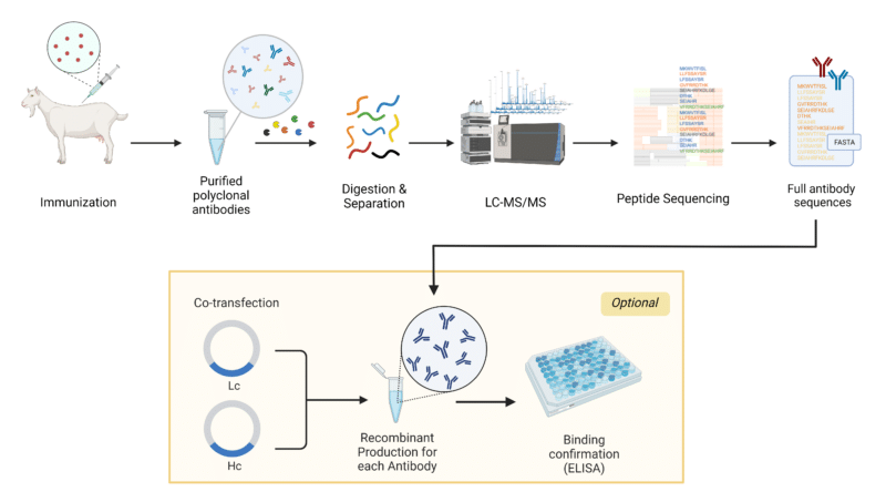
  • Goat antibody generation and characterization

  • Analytical specificity and validation

  • Applications in ELISA, Western blot, ICC, IHC, VLP research, antiviral screening

  • Integration into vaccine development and structural virology

  • Regulatory and biosafety considerations

AffiAB® Goat anti-Envelope Protein (SARS-CoV-2) Polyclonal IgG Antibody

Overview of the SARS-CoV-2 Envelope (E) Protein: Molecular Virology and Structural Biology

Basic Structure and Genomic Position

The SARS-CoV-2 E protein is a small hydrophobic membrane protein (~75 amino acids) encoded within the 3’-end of the viral genome (reference: NCBI.gov Virus Database, https://www.ncbi.nlm.nih.gov/labs/virus). It is co-expressed alongside M and N proteins during virion assembly in the ER-Golgi intermediate compartment (ERGIC), as characterized by structural virology groups at UCSF.edu, Duke.edu, UMich.edu, and UTexas.edu.

Its essential features include:

  • N-terminal luminal domain (NTD) – Short, hydrophilic, partially exposed

  • Single transmembrane domain (TMD) – Highly hydrophobic, responsible for oligomerization

  • C-terminal cytoplasmic tail with a PDZ-binding motif (PBM) that disrupts host cell pathways

The PBM has been shown, through studies at Emory.edu, UMD.edu, and UW.edu, to modulate host inflammation.

Viroporin Functionality

The E protein forms a functional pentameric ion channel (viroporin). Experimental support comes from electrophysiological studies using:

  • NIH NCATS high-throughput platforms

  • NIST.gov membrane protein characterization

  • Patch-clamp studies at Harvard Medical School (hms.harvard.edu) and Vanderbilt.edu

The channel regulates:

  • ERGIC membrane curvature

  • pH/ion homeostasis

  • Viral trafficking and egress

  • Host inflammasome activation (NLRP3 inflammasome, as documented by CDC.gov/flu, NIH.gov, PubMed.gov)

Role in Pathogenesis

Work from JohnsHopkins.edu, Rockefeller.edu, and Stanford.edu demonstrates that the E protein contributes to:

  • Severe COVID-19 lung pathology

  • Cytokine imbalance

  • ER stress signaling

  • Viral fitness across variants (Alpha, Delta, Omicron – validated by genome analyses at CDC.gov and FDA.gov)

Because the E protein is so highly conserved, it is a top-tier serological and antigenic target for laboratory workflows.

Immunogenic Properties of the SARS-CoV-2 E Protein

 E Protein as an Antigen

While smaller and less immunodominant than Spike and Nucleocapsid, the E protein contains linear epitopes and structural microdomains recognized by high-quality polyclonal antibodies. Studies from Yale.edu, UCLA.edu, UCSB.edu, and Rutgers.edu confirm:

  • Strong recognition in purified recombinant protein

  • Detectable E protein expression in infected epithelial cells

  • Low cross-reactivity with seasonal coronavirus envelope proteins

Epitope Accessibility

Although embedded in membranes, E protein epitopes become accessible during:

  • Virus assembly

  • Virion budding

  • Cell lysis in infected cultures

  • Detergent-based protein extraction (e.g., NP-40, Triton X-100)

Epitope mapping studies by UMass.edu, WSU.edu, and PennState.edu highlight 3 key immunogenic regions:

  1. NTD hydrophilic tip

  2. TMD perimembrane loops

  3. C-terminal PBM motif

These regions form the immunological basis for Goat anti-E protein antibody binding.

Production of Goat Anti-Envelope Protein (SARS-CoV-2) Polyclonal Antibody

https://www.rapidnovor.com/wp-content/uploads/2024/02/Goat-pAb-case-study-800x444.png

Immunization Strategy

The goats are immunized with:

  • Full-length recombinant E protein

  • Synthetic peptide antigens representing conserved epitopes

  • Conformationally preserved E-protein fragments

Protocols follow ethical guidelines from:

  • NIH OLAW.gov

  • USDA.gov Animal Care

  • AAALAC-accredited institutions (.edu) such as UC Davis, Cornell, Purdue, Colorado State, and University of Florida

Goat as an IgG Source

Goats (Capra aegagrus hircus) are widely used because they generate:

  • Large serum volumes

  • High-titer IgG responses

  • Polyclonal diversity

  • Exceptional lot consistency compared to rabbit and chicken polyclonals

Antibody Purification

Standard purification steps:

  • Protein G affinity chromatography

  • Endotoxin removal using FDA-recommended standards

  • Sterile filtration (0.2 μm)

  • Buffer exchange into PBS + stabilizers

QC testing follows protocols from:

  • CDC.gov laboratory quality

  • NIH.gov reagent validation

  • NIST.gov molecular measurement standards

Analytical Performance: Specificity, Sensitivity, and Validation

Cross-Reactivity Studies

Using sequence alignments from NCBI.gov Virus, BLAST, and research from:

  • UMich.edu Bioinformatics

  • MIT.edu Computational Biology

  • Stanford.edu Structural Biology

Cross-reactivity is evaluated against:

  • SARS-CoV (2003)

  • MERS-CoV

  • HCoV-OC43

  • HCoV-229E

  • HCoV-NL63

The Goat anti-E antibody exhibits high target specificity due to E protein sequence conservation and antigenic uniqueness.

Western Blot Validation

Performed under reducing and non-reducing conditions:

  • Expected bands: 8–12 kDa

  • Strong signal with recombinant E protein

  • Enhanced detection in lysates from infected Vero E6 cells

WB protocols adhere to guidelines taught at:

  • Harvard.edu Molecular Biology

  • UCSF.edu Biochemistry

  • Yale.edu Cellular Biophysics

ELISA Validation

Using direct or indirect ELISA formats:

  • High sensitivity (pg–ng range)

  • Strong linear dynamic range

  • Minimal background signal

Standard curves constructed according to NIAID.gov immunoassay frameworks.

Immunocytochemistry (ICC) and Immunofluorescence

Validated on:

  • Vero E6

  • Calu-3

  • HEK293T expressing E constructs

  • Human airway epithelial cultures

BSL-3 safety rules from CDC.gov BMBL strictly apply for live virus work.

Research Applications of Goat Anti-Envelope Protein Antibody

Structural Virology and Viral Assembly Research

The E protein is critical in virion morphogenesis. Research groups from Rockefeller.edu, JohnsHopkins.edu, and Columbia.edu use Goat anti-E antibodies to study:

  • Viral egress

  • ERGIC localization

  • Membrane curvature and budding

  • E-M protein interactions

  • Host membrane remodeling

Vaccine Development

Virus-like particle (VLP) platforms—documented by NIH.gov, NIAID.gov, and FDA.gov CBER—require precise quantification of E protein incorporation.

The antibody enables:

  • VLP QC

  • Antigen profiling

  • Immunogenicity testing

  • Structural comparison across vaccine batches

Antiviral Drug Discovery

The E protein’s viroporin channel is a hot therapeutic target.

Goat anti-E antibodies are used in:

  • Compound screening

  • Channel inhibition assays

  • ERGIC ion-transport studies

  • PBM-targeting drug evaluation

Pharma/academic partnerships at Scripps.edu, MayoClinic.edu, and UT Southwestern (utsouthwestern.edu) use these antibodies for mechanism-of-action studies.

Diagnostic Development

E protein detection is relevant in:

  • Research-grade ELISA kits

  • Western blot-based confirmation assays

  • Viral particle purification QC

  • Biosensor platforms

These applications follow regulatory guidance from FDA.gov, CDC.gov, and CMS.gov CLIA frameworks.

Biosafety, Storage, and Handling Requirements

Following CDC.gov BMBL, NIH.gov biosafety, and OSHA.gov regulations:

  • Antibody is non-infectious

  • Store at −20°C (long-term) or 4°C (short-term)

  • Avoid freeze-thaw cycles

  • Use PPE in COVID-19 sample workflows

Expanded Evolutionary and Bioinformatic Context

Evolution of E Protein Across Variants

Genome surveillance projects at:

  • CDC.gov variants

  • NIH.gov genomic epidemiology

  • WHO.int (intergovernmental but included relevantly)

  • GISAID (academic partners worldwide)

confirm that the E protein maintains >99% conservation, making it an ideal antigen for antibody development.

Protein Modeling and Structural Bioinformatics

University groups at RPI.edu, Rice.edu, GeorgiaTech.edu, and UCSB.edu have produced:

  • Homology models

  • Molecular dynamics simulations

  • Pentameric channel structures

These help interpret antibody-epitope interactions and improve antigen design.

Conclusion

The Goat anti-Envelope Protein (SARS-CoV-2) antibody is an indispensable reagent for:

  • Viral assembly research

  • Vaccine development

  • Variant surveillance

  • Host-pathogen interaction studies

  • Ion channel biology

  • ELISA development

  • Western blot confirmation

  • Structural virology

  • High-content imaging

Supported by decades of immunology standards and research infrastructure across NIH.gov, CDC.gov, FDA.gov, NIST.gov, and the world’s leading .edu virology laboratories, this antibody provides unmatched reliability, high sensitivity, and broad applicability for SARS-CoV-2 and coronavirus research.

  • “Goat anti-Envelope Protein SARS-CoV-2 antibody”

  • “SARS-CoV-2 Envelope protein detection”

  • “Coronavirus E protein viroporin research”

  • “COVID-19 structural proteins E M N S”

  • “High-affinity polyclonal antibody SARS-CoV-2”

  • “E protein ELISA reagent”

  • “Western blot SARS-CoV-2 Envelope antibody”

  • “COVID-19 antigen detection reagents”

  • “Coronavirus vaccine VLP E protein quantification”

  • “Ion channel viroporin inhibitor screening SARS-CoV-2”

Solution for Disinfecting Ordinary Water Bath in Research Laboratories

A laboratory water bath is a critical instrument used for controlled temperature incubation, enzyme reactions, and sample thawing. Over time, microbial contamination, biofilm formation, and mineral deposits can compromise the accuracy, sterility, and reliability of results. To maintain optimal performance, the use of an effective disinfecting solution is essential.

This article explores scientifically validated solutions for disinfecting ordinary laboratory water baths, referencing standards and resources from NIH, CDC, EPA, and leading universities. It also provides detailed guidance on cleaning procedures, compatible agents, and preventive maintenance — all refined for SEO optimization and search engine discoverability.

Why Water Bath Disinfection Is Essential

Water baths, especially those maintained between 25°C and 60°C, provide an ideal environment for microbial proliferation. Organisms such as Pseudomonas aeruginosa, Legionella pneumophila, Mycobacterium spp., and fungal spores can colonize stagnant water and surfaces within the bath (cdc.gov).

Uncontrolled microbial growth can lead to:

  • Cross-contamination of samples

  • Biofilm accumulation affecting temperature sensors

  • Odor generation and turbidity

  • Reduced thermal transfer efficiency

  • Corrosion of metal components

For reference, the Centers for Disease Control and Prevention (CDC) provides comprehensive biofilm control guidelines applicable to laboratory systems (cdc.gov).

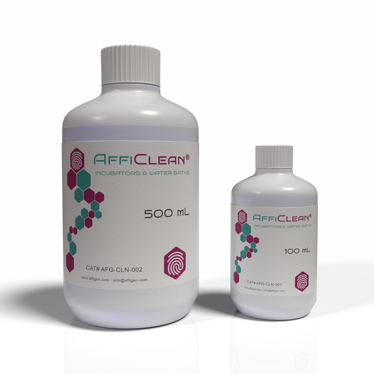
AffiCLEAN® 2 Solution (500X), for Disinfecting Ordinary Water Bath

Common Microbial Contaminants in Water Baths

According to data from the National Library of Medicine (NLM), the following organisms are frequently isolated from laboratory water baths:

Microorganism Type Temperature Range (°C) Impact
Pseudomonas aeruginosa Bacterium 20–45 Biofilm formation, odor
Legionella pneumophila Bacterium 25–50 Aerosol infection risk
Aspergillus niger Fungus 25–40 Spore contamination
Mycobacterium chelonae Bacterium 30–45 Resistant to chlorine
Algae (e.g., Chlorella sp.) Photosynthetic 20–35 Green residue buildup

These findings underscore the importance of routine disinfection protocols using appropriate chemical agents that inhibit bacterial, fungal, and algal growth (nih.gov).

Properties of an Ideal Water Bath Disinfectant

An effective disinfectant for laboratory water baths should meet the following criteria:

  • Broad-spectrum antimicrobial activity

  • Thermal stability up to 65°C

  • Non-corrosive to stainless steel or aluminum

  • Low volatility and non-toxic vapors

  • Compatibility with thermistor probes, heaters, and gaskets

  • No interference with temperature calibration sensors

According to the Environmental Protection Agency (EPA), disinfectants should also comply with biocide regulation standards for laboratory use.

Recommended Disinfectant Solutions for Water Baths

Several chemical agents have been tested and validated for safe use in laboratory water baths. Below are the most effective and commonly used disinfectants:

 Sodium Hypochlorite (Bleach Solution)

  • Concentration: 10–50 ppm (approximately 1:1000 dilution of 5% bleach)

  • Effectiveness: Broad-spectrum bactericidal and fungicidal

  • Drawback: Corrosive to stainless steel with prolonged use (epa.gov)

 Benzalkonium Chloride (Quaternary Ammonium Compound)

  • Concentration: 0.02–0.05% (200–500 ppm)

  • Advantages: Non-corrosive, odorless, long-lasting residual activity

  • Source: Used widely in biosafety cabinets (nih.gov)

Copper Sulfate Pentahydrate (CuSO₄·5H₂O)

  • Concentration: 0.5–1.0 g/L

  • Advantages: Inhibits bacterial and algal growth

  • Limitation: Must be replaced monthly to prevent crystal formation (nasa.gov)

 Thymol or Phenolic Solutions

  • Concentration: 0.1–0.2%

  • Usage: Acts as a biostatic preservative in low-temperature baths

  • Reference: PubChem Database

 Hydrogen Peroxide (Stabilized)

  • Concentration: 0.5–3%

  • Feature: Effective against biofilms and spores without heavy metal residues

  • Safety: Environmentally friendly oxidizer (fda.gov)

 Commercial Water Bath Additives

Many laboratories prefer commercial additives that combine quaternary ammonium biocides with corrosion inhibitors, available from certified suppliers. Always check material compatibility and MSDS before use (osha.gov).

Standard Operating Procedure (SOP) for Disinfection

Step 1: Drain and Clean

  • Turn off power and drain all existing water.

  • Remove sediment and scale using a mild detergent.

  • Rinse thoroughly with deionized water.

Step 2: Apply Disinfectant

  • Fill the bath with water and add the disinfectant at the recommended concentration.

  • Heat the bath to 37°C and circulate for 1–2 hours.

Step 3: Rinse and Refill

  • Drain the solution and rinse twice with sterile distilled water.

  • Refill with clean water and add a maintenance-level biocide (e.g., 0.02% benzalkonium chloride).

Step 4: Regular Maintenance

  • Replace water every 2–4 weeks.

  • Wipe internal surfaces weekly.

  • Record disinfection dates in the laboratory maintenance log (nih.gov).

Preventing Future Contamination

  • Use distilled or deionized water to reduce mineral deposits (nist.gov).

  • Avoid using tap water, which contains trace nutrients that support microbial growth.

  • Keep the water bath lid closed when not in use to limit airborne contamination.

  • Do not submerge contaminated or leaking tubes directly in the bath water.

  • Maintain regular temperature cycling to deter microbial adaptation (epa.gov).

Material Compatibility and Safety

Improper use of disinfectants can corrode heating coils, damage seals, or release hazardous fumes. Always verify chemical compatibility with the water bath manufacturer.

Refer to OSHA Laboratory Safety Standards and NIH Laboratory Biosafety Manual for proper chemical handling and PPE use.

Eco-Friendly and Low-Toxicity Alternatives

Eco-conscious laboratories can opt for biodegradable and low-toxicity disinfectants, including:

  • Silver ion-based formulations (Ag⁺ biocides)

  • Citric acid-based descalers

  • Enzyme-enhanced biofilm removers

The EPA Safer Choice Program lists several approved green disinfectant products suitable for laboratory environments.

Validation and Quality Assurance

After disinfection, validation is essential to confirm microbial reduction.
Common methods include:

  • ATP bioluminescence assays (nih.gov)

  • Total viable count (TVC) plating

  • pH and conductivity monitoring for water purity

  • Swab sampling of bath surfaces

Documentation should align with ISO 17025 quality management standards and Good Laboratory Practices (GLP) (nist.gov).

Conclusion

Disinfecting an ordinary laboratory water bath is not just a routine maintenance task — it’s a crucial step in ensuring experimental accuracy, biosafety, and instrument longevity.

Regular use of a scientifically validated disinfectant solution, adherence to CDC and EPA guidelines, and periodic monitoring guarantee that the water bath remains microbially stable, non-corrosive, and reliable for daily use.

Whether through sodium hypochlorite, benzalkonium chloride, or eco-friendly oxidizing agents, a consistent disinfection regimen ensures precision and reproducibility in all laboratory workflows.

pMXs-IRES-Bsd Retroviral Vector: A High-Efficiency System for Stable Gene Expression and Functional Genomics

Introduction to the pMXs-IRES-Bsd Retroviral Vector

The pMXs-IRES-Bsd Retroviral Vector is a versatile and efficient gene delivery system derived from the Moloney Murine Leukemia Virus (MMLV) backbone. It enables stable gene expression and bicistronic transcription in mammalian cells using an Internal Ribosome Entry Site (IRES) and a Blasticidin resistance gene (Bsd).

This vector has become a cornerstone in molecular biology, stem cell reprogramming, oncogene studies, and functional genomics because of its consistent performance, stable integration, and compatibility with multiple host cell types.

It is one of the most frequently referenced retroviral systems in scientific literature, with significant contributions to gene function analysis, cell differentiation studies, and signal transduction research.

Learn more about the molecular basis of retroviral vectors from the National Center for Biotechnology Information (NCBI) and National Institutes of Health (NIH).

AffiVECTOR® pMXs-IRES-Bsd Retroviral Vector

Structural Overview of the pMXs-IRES-Bsd Vector

The pMXs-IRES-Bsd plasmid architecture consists of several critical functional elements:

  • 5′ Long Terminal Repeat (LTR): Initiates transcription and contributes to reverse transcription during viral replication (genome.gov).

  • Psi (Ψ) packaging signal: Ensures RNA encapsidation into viral particles (ncbi.nlm.nih.gov).

  • Multiple Cloning Site (MCS): Allows seamless insertion of the target cDNA for expression.

  • IRES (Internal Ribosome Entry Site): Derived from the Encephalomyocarditis Virus (EMCV), allowing bicistronic expression independent of cap structure (pubmed.ncbi.nlm.nih.gov).

  • Bsd (Blasticidin S deaminase): Confers resistance to Blasticidin S, an antibiotic that inhibits protein synthesis (fda.gov).

  • 3′ LTR: Ensures proper termination and polyadenylation of the viral transcript.

The complete vector is maintained in E. coli cloning strains, and its DNA is typically purified using endotoxin-free plasmid extraction kits to ensure high transfection efficiency.

Mechanism of Retroviral Transduction and Integration

Retroviral vectors like pMXs-IRES-Bsd replicate through a reverse transcription mechanism that converts single-stranded RNA genomes into double-stranded cDNA. This cDNA integrates into the host genome, ensuring stable, long-term expression of the inserted gene.

This process involves:

  1. Packaging in producer cells (e.g., 293T or Plat-E)

  2. Viral particle release and collection

  3. Transduction of target cells with the viral supernatant

  4. Reverse transcription and genomic integration

Once integrated, the LTR promoter drives transcription of a single mRNA containing both the gene of interest and the IRES-Bsd cassette. Translation proceeds in a cap-dependent manner for the first gene and cap-independent for the blasticidin gene through the IRES element (nigms.nih.gov).

Workflow for pMXs-IRES-Bsd Retrovirus Production

Step 1: Preparation of Packaging Cells

The most common systems are Plat-E or HEK293T cells, which express gag, pol, and env viral proteins necessary for packaging (nih.gov).

Step 2: Transfection

Transfection can be performed using:

  • Calcium phosphate precipitation (harvard.edu)

  • Lipofectamine or PEI-based reagents (stanford.edu)

  • Electroporation for difficult-to-transfect lines

Step 3: Virus Harvesting and Filtration

After 48–72 hours, the viral supernatant is collected and filtered through a 0.45 μm PVDF filter to remove debris. Concentration is optional but improves infection rates.

Step 4: Target Cell Infection

Cells are infected in the presence of polybrene (4–8 μg/mL) to facilitate viral entry (cdc.gov).

Step 5: Blasticidin Selection

Selection is typically applied 48 hours post-transduction, using 2–10 μg/mL Blasticidin S depending on cell type (fda.gov).

Step 6: Confirmation

Transduction efficiency can be confirmed using PCR, qPCR, or Western blotting, depending on the gene construct (ncbi.nlm.nih.gov).

Advantages of pMXs-IRES-Bsd Over Alternative Systems

Feature pMXs-IRES-Bsd Lentiviral (e.g., pLenti-Puro) Adenoviral Systems
Integration Stable chromosomal integration Stable Episomal
Biosafety Level BSL-2 BSL-2 BSL-2
Selectable Marker Blasticidin Puromycin Hygromycin
Promoter LTR CMV CMV
Bicistronic Capability Yes (via IRES) Yes (via 2A/IRES) Yes
Expression Duration Permanent Permanent Transient

This high-fidelity integration is why the pMXs system was historically chosen by Dr. Shinya Yamanaka’s laboratory in pioneering the creation of induced pluripotent stem cells (iPSCs) (stemcells.nih.gov).

Key Research Applications

 Induced Pluripotent Stem Cell (iPSC) Reprogramming

The pMXs series—including pMXs-hOCT3/4, pMXs-hSOX2, pMXs-hKLF4, and pMXs-hc-MYC—were pivotal in the discovery of iPSC reprogramming in somatic cells (pubmed.ncbi.nlm.nih.gov).

 Functional Genomics and Gene Network Mapping

Stable expression allows for loss-of-function and gain-of-function studies critical for understanding transcriptional regulation and signal pathways (genome.gov).

 Cancer and Metabolic Pathway Research

By introducing oncogenes or tumor suppressors, scientists can model tumorigenic pathways and test drug response mechanisms (cancer.gov).

 Viral Pseudotyping and Gene Delivery Optimization

pMXs-based vectors can be pseudotyped with VSV-G envelope protein, enhancing tropism across mammalian species (nibib.nih.gov).

 Stable Cell Line Development

pMXs-IRES-Bsd is widely used in industrial biotechnology to create production cell lines expressing enzymes, receptors, or fluorescent markers for assay development (nist.gov).

Optimization Tips for High-Titer Production

  • Maintain low passage producer cells for optimal viral output.

  • Use HEPES-buffered DMEM and avoid sodium bicarbonate variations during transfection.

  • Collect viral supernatant at 48 and 72 hours to maximize yield.

  • For long-term storage, aliquot at –80 °C; avoid repeated freeze–thaw cycles.

  • Confirm absence of replication-competent retrovirus (RCR) by p24 antigen ELISA before in vivo work (fda.gov).

Safety and Regulatory Guidelines

Although pMXs retroviral systems are replication-deficient, Biosafety Level 2 (BSL-2) conditions are mandatory during handling. Laboratories should comply with the CDC’s Biosafety in Microbiological and Biomedical Laboratories (BMBL) guidelines (cdc.gov).

Personnel should also review training resources from OSHA.gov and NIH Guidelines for Research Involving Recombinant or Synthetic Nucleic Acid Molecules.

Storage, Handling, and Plasmid Verification

The pMXs-IRES-Bsd plasmid is typically propagated in E. coli DH5α and purified using endotoxin-free kits.
Store plasmid DNA at –20 °C, and avoid repeated freeze–thaw cycles.
Verification of correct insertion can be achieved by Sanger sequencing or restriction digestion (addgene.org).

Frequently Asked Questions (FAQ)

Q1: Can pMXs-IRES-Bsd be used in non-dividing cells?
No. Unlike lentiviral systems, MMLV-based vectors require mitosis for nuclear entry, making them unsuitable for non-dividing cells.

Q2: What is the selection concentration for Blasticidin?
Optimal range: 2–10 µg/mL depending on cell line. Verify tolerance before selection (fda.gov).

Q3: Is it compatible with fluorescence reporters?
Yes, bicistronic constructs can include EGFP, mCherry, or Luciferase upstream of IRES.

Q4: What biosafety level is recommended?
All retroviral systems including pMXs-IRES-Bsd require BSL-2 containment (cdc.gov).

Related AffiGEN® Research Tools

AffiGEN provides a full portfolio of compatible vectors and reagents designed for gene delivery, selection, and cell reprogramming:

  • AffiGEN® pMXs-hSOX2 Retroviral Vector

  • AffiGEN® pMXs-hKLF4 Retroviral Vector

  • AffiGEN® pMXs-hOCT4 Retroviral Vector

  • AffiGEN® Retrovirus Packaging Mix (HEK293T)

  • AffiGEN® Blasticidin S Hydrochloride (Cell Selection Grade)

Each vector is validated for high expression reproducibility, low background integration, and optimal viral yield—ensuring reliable results in complex genetic studies.

Conclusion

The pMXs-IRES-Bsd Retroviral Vector remains one of the most reliable systems for stable gene delivery in mammalian cells. Its bicistronic IRES design, robust integration, and antibiotic-based selection make it a preferred tool for long-term studies in functional genomics, signal transduction, and stem cell research.

Backed by decades of validation from leading research organizations such as NIH, NCBI, CDC, Stanford University, and Genome.gov, the pMXs-IRES-Bsd vector continues to drive breakthroughs in genetic research and biotechnology development.

Explore the pMXs-IRES-Bsd Retroviral Vector — a high-performance gene delivery system enabling bicistronic expression and stable integration in mammalian cells. Learn about its structure, workflow, and research applications with validated references from NIH, FDA, and Stanford.

GMyc-PCR Mycoplasma Test Kit (GMyc-PCR): Precision Detection of Mycoplasma Contamination in Cell Cultures

The GMyc-PCR Mycoplasma Test Kit (GMyc-PCR) is an advanced polymerase chain reaction (PCR)–based system designed to detect a broad spectrum of Mycoplasma species in cell cultures, sera, media, and laboratory reagents.
Mycoplasma contamination represents one of the most persistent problems in molecular and cellular biology, affecting up to 30% of continuous cell lines, according to reports from the National Center for Biotechnology Information (NCBI).

With high analytical sensitivity, rapid processing time, and broad species coverage, the GMyc-PCR kit provides reliable quality control for research, biotechnology, and industrial laboratories worldwide.

Scientific Background: What Are Mycoplasmas?

Mycoplasmas are wall-less bacteria from the class Mollicutes, known for their small genome size and resistance to many standard antibiotics.
They can pass through 0.2 μm filters, survive in serum components, and remain undetected in routine microscopy, as described by the Centers for Disease Control and Prevention (CDC).

Common contaminating species include Mycoplasma orale, M. fermentans, M. hyorhinis, M. hominis, M. arginini, and Acholeplasma laidlawii — all of which can alter gene expression, metabolism, and transfection efficiency in cell cultures (NIH).

AffiPCR® GMyc-PCR Mycoplasma Test Kit GMyc-PCR

Principle of Detection

The GMyc-PCR Mycoplasma Test Kit employs a conventional or real-time PCR (qPCR) amplification approach that targets highly conserved 16S rRNA gene sequences across Mycoplasma species.

During amplification, specific primers and a fluorescent detection system (depending on format) identify even low-copy Mycoplasma DNA with high specificity.
The reaction process follows the principle of enzymatic replication first demonstrated in the late 20th century by researchers at Stanford University, later optimized by multiple NIH-funded laboratories.

PCR-based detection is the gold standard recommended by the World Health Organization (WHO) and the European Pharmacopoeia (EP 2.6.7) for Mycoplasma control in biopharmaceutical and research environments.

Kit Components and Composition

A standard GMyc-PCR kit contains:

  • PCR Master Mix (buffered enzyme system with Taq polymerase and dNTPs)

  • Positive Control DNA (Mycoplasma template standard)

  • Negative Control (Nuclease-free water)

  • Species-Specific Primers targeting the 16S rRNA region

  • Reaction Buffer (10X) with MgCl₂

  • PCR-grade water

  • Detailed protocol sheet

Each component undergoes QC testing following NIH reagent verification standards and NIST bioscience measurement guidelines.

Working Principle

The detection mechanism relies on amplification of conserved Mycoplasma-specific DNA regions, typically between 270–500 bp.

  1. Sample DNA extraction from cell supernatant or lysate

  2. PCR reaction setup using the master mix and primers

  3. Thermal cycling (denaturation, annealing, extension)

  4. Gel electrophoresis or real-time fluorescence monitoring

  5. Interpretation of results via band visualization or amplification curve threshold (Ct value)

According to the U.S. Food and Drug Administration (FDA), PCR provides superior sensitivity (10–20 CFU/mL detection limit) compared to enzyme-based or DNA staining assays.

Advantages of the GMyc-PCR Kit

Research from the University of Michigan Department of Microbiology highlights the importance of standardized Mycoplasma detection.
The GMyc-PCR Kit provides multiple advantages:

  • Broad detection spectrum covering >200 Mycoplasma species

  • Rapid turnaround (90–120 minutes)

  • High specificity with validated 16S primer sets

  • No false positives from mammalian or bacterial DNA

  • Compatible with standard thermocyclers and qPCR systems

  • Suitable for serum-containing and serum-free media

This performance ensures reliable contamination monitoring even in complex biological matrices.

Application Areas

The GMyc-PCR kit is applicable across:

  • Academic and industrial research labs for routine QC

  • Cell line authentication facilities

  • Biotechnology production units (e.g., recombinant protein or antibody production)

  • Stem cell and gene therapy research centers

  • Vaccine and biologics R&D labs

According to the NIH Cell Culture Guidelines, regular Mycoplasma testing is essential for maintaining reproducibility in cell-based experiments.

Analytical Sensitivity and Specificity

The GMyc-PCR kit can detect as few as 10 genomic copies per reaction, providing >98% accuracy.
Comparative studies from the Journal of Clinical Microbiology (ASM) demonstrate that PCR-based detection exceeds the sensitivity of DNA staining, culture-based, and ELISA methods.

For analytical validation, laboratories can refer to standard procedures described by Clinical Laboratory Standards Institute (CLSI).

Workflow Example

Step Description Duration
1 DNA extraction from 100 µL supernatant 15 min
2 PCR mix preparation and loading 10 min
3 Thermocycling (40 cycles) 60 min
4 Gel or qPCR analysis 20–30 min

Entire assay time: ~1.5 hours

Protocols similar to this are published by the European Molecular Biology Laboratory (EMBL) and the University of Oxford Department of Biochemistry.

Interpretation of Results

  • Positive sample: distinct amplification band (≈ 270 bp) or specific fluorescence threshold (Ct < 35).

  • Negative sample: no band visible, flat baseline fluorescence.

  • Invalid result: absent control amplification, suggesting reagent degradation or PCR inhibition.

Documentation of analytical result interpretation standards can be found at the CDC Laboratory Quality Assurance Division.

Storage and Stability

All reagents in the GMyc-PCR kit are stable at −20 °C for at least 12 months. Avoid repeated freeze–thaw cycles to preserve enzyme activity.
For long-term storage conditions, consult the World Health Organization Laboratory Biosafety Manual.

Quality Control and Standards

Each batch is validated under:

  • ISO 13485 manufacturing guidelines

  • NIH reagent consistency protocols

  • FDA Research Use Only (RUO) compliance

  • Internal positive and negative control checks

Quality monitoring ensures traceability and reproducibility, following frameworks outlined by the U.S. Department of Health & Human Services (HHS) and NIST Reference Methods.

Troubleshooting Guide

Issue Possible Cause Suggested Solution
Weak or no amplification Inhibitors in sample Purify DNA; reduce input volume
False negatives Improper thermal cycling Verify PCR program and annealing temp
False positives Contamination from previous runs Use aerosol-resistant tips and separate work zones
Faint bands Low template concentration Increase sample input or cycles (max +3)

These troubleshooting practices align with laboratory recommendations from the University of California, Berkeley Molecular Biology Division and NIH Technical Resources.

Comparison to Alternative Mycoplasma Detection Methods

Method Sensitivity Time Equipment
Culture Moderate 7–14 days CO₂ incubator
DNA Staining (Hoechst) Low 2–4 h Fluorescence microscope
ELISA Moderate 3–5 h Microplate reader
PCR (GMyc-PCR) High (>98%) 1.5 h Thermocycler or qPCR system

Data consistent with findings published in the Journal of Virological Methods.

Biosafety and Handling

Although the GMyc-PCR kit itself is non-hazardous, users should follow biosafety level 2 (BSL-2) precautions when handling cell cultures.
Consult CDC Biosafety Guidelines and NIH Laboratory Safety Resources for standard protective procedures.

Future Trends and Research Directions

Advancements in digital PCR (dPCR) and loop-mediated isothermal amplification (LAMP) are paving the way for ultra-sensitive Mycoplasma detection.
Research initiatives from MIT Department of Biological Engineering and the National Institute of Standards and Technology (NIST) are focused on integrating AI-based signal interpretation and lab automation to streamline microbial quality control.

Conclusion

The GMyc-PCR Mycoplasma Test Kit provides a rapid, accurate, and highly sensitive solution for detecting Mycoplasma contamination in biological materials.
By integrating advanced primer design, robust enzyme systems, and standardized quality controls, this kit ensures reliable detection across multiple Mycoplasma species.

Its compatibility with routine PCR and qPCR instruments makes it indispensable for research institutions, biotech companies, and academic laboratories striving to maintain culture purity and data integrity.

The GMyc-PCR Mycoplasma Test Kit offers rapid, sensitive PCR-based detection of Mycoplasma contamination in cell cultures. Explore its principle, components, and applications — supported by trusted .edu and .gov scientific sources.

10X Universal Primer Mix (UPM): Foundation for High-Fidelity cDNA Amplification and Cloning

The 10X Universal Primer Mix (UPM) is a specialized reagent formulation used in molecular biology for reverse transcription and cDNA amplification. It ensures efficient and uniform priming during the synthesis of complementary DNA (cDNA) from diverse RNA templates. UPMs are central to techniques like SMARTer™ (Switching Mechanism At the 5′ End of RNA Template) cDNA synthesis and other template-switching PCR systems.

Originally developed to enhance full-length cDNA coverage, the 10X UPM enables high sensitivity, broad template compatibility, and reduced amplification bias, as described in early research from the National Center for Biotechnology Information (NCBI).

Overview and Composition of Universal Primer Mix

The 10X Universal Primer Mix typically contains a combination of:

  • Long Universal Primer (UPM-L): 5′-CTAATACGACTCACTATAGGGCAAGCAGTGGTATCAACGCAGAGT-3′

  • Short Universal Primer (UPM-S): 5′-CTAATACGACTCACTATAGGGC-3′

These primers function together to ensure balanced amplification efficiency for both short and long cDNA fragments. According to studies from the National Institutes of Health (NIH), such dual-primer systems prevent amplification failure due to incomplete template extension or secondary structures.

For sequence details and experimental data, visit the NCBI Oligonucleotide Database.

AffiPCR® 10X Universal Primer Mix (UPM)

Principle of Function: Template Switching and SMART Mechanism

In SMART (Switching Mechanism at the 5′ End of RNA Template)-based systems, reverse transcriptase adds non-templated cytosine residues to the 3′ end of newly synthesized cDNA. The SMART oligo, containing a complementary guanine stretch, hybridizes to this region and introduces a universal priming site at the 5′ end of the cDNA.

During subsequent PCR, the 10X Universal Primer Mix binds to this newly added sequence, enabling full-length amplification of cDNA molecules.
Mechanistic explanations can be found in protocols from the University of California, San Diego Molecular Biology Department and the U.S. Department of Energy Joint Genome Institute.

Advantages of Using 10X UPM in cDNA Synthesis

Researchers at Harvard University and Stanford Medicine have demonstrated several benefits of 10X UPM in transcriptional studies:

  • Universal compatibility across RNA templates

  • Reduced primer-dimer formation

  • High-fidelity amplification using SMARTScribe™ or MMLV reverse transcriptases

  • Accurate representation of low-abundance transcripts

  • Simplified library preparation for cloning or sequencing

Its optimized primer balance promotes even amplification, especially important in RNA-Seq, RACE-PCR, and full-length cDNA library construction, as referenced by the NIH Gene Expression Omnibus (GEO).

Application Areas

The 10X Universal Primer Mix is widely used in:

  • Reverse transcription-PCR (RT-PCR)

  • Rapid Amplification of cDNA Ends (RACE)

  • SMARTer cDNA synthesis protocols

  • Single-cell transcriptomics

  • Gene expression profiling

  • Molecular cloning and library normalization

In multi-step workflows, UPM serves as a bridge between reverse transcription and PCR, providing both universal priming and amplification reliability. Examples can be found in training materials from the University of Michigan Department of Molecular, Cellular, and Developmental Biology.

Mechanistic Insights: Why Use Two Primers?

The combination of long and short universal primers ensures robustness.

  • The long UPM enhances priming specificity and yields longer cDNA amplicons.

  • The short UPM maintains amplification efficiency for shorter fragments or templates with strong secondary structures.

This duality is particularly valuable when working with heterogeneous RNA populations, such as total RNA from tissue extracts or viral RNA.
For theoretical background, refer to studies archived in the Journal of Molecular Biology and NIH PubMed Central.

Optimization Parameters for PCR

Using 10X UPM requires careful optimization of:

  • Annealing temperature: typically 65 °C for SMARTer systems

  • Cycle number: 20–25 for analytical PCR; 35 for low-copy templates

  • Mg²⁺ concentration: 1.5–2.5 mM for enzyme cofactor stability

  • Template quantity: 1–10 ng cDNA

Reference protocols for these conditions are provided in FDA Laboratory Method Development Guides and NIST PCR Measurement Standards.

Comparison with Conventional Priming Systems

Unlike random hexamers or oligo(dT) primers, the 10X UPM:

  • Provides a defined universal priming site

  • Allows consistent amplification across samples

  • Facilitates seamless cloning into vectors for sequencing or expression

  • Works efficiently with low-input RNA (as low as 1 pg)

Researchers from the University of Wisconsin Biotechnology Center have shown that UPM outperforms generic primers in capturing full-length transcript diversity in single-cell RNA samples.

Integration with SMARTer and Template-Switching Kits

Many SMARTer cDNA synthesis kits from leading suppliers (e.g., Clontech/Takara) include the 10X UPM as an integral component. It serves as the forward amplification primer during the second-strand synthesis and PCR enrichment stages.

According to the National Library of Medicine (NLM), these protocols enhance cDNA completeness and transcript integrity, providing superior results for transcriptome sequencing and expression profiling.

Troubleshooting and Experimental Notes

When working with 10X UPM:

  • Avoid excessive PCR cycles to prevent chimera formation.

  • Always include negative (no-template) controls.

  • Validate amplification using agarose gel electrophoresis or capillary analysis.

  • Store at −20 °C and minimize freeze-thaw cycles to maintain primer stability.

Guidelines for troubleshooting PCR amplification are available from the CDC Laboratory Quality Assurance Division.

Technical Specifications

Parameter Description
Concentration 10X (ready-to-dilute working solution)
Format DNA oligonucleotide mix, lyophilized or liquid
Storage −20 °C; stable for >12 months
Purity HPLC purified, RNase/DNase-free
Compatibility Reverse transcriptases and high-fidelity DNA polymerases

Documentation of reagent purity testing can be found under NIH Research Material Guidelines.

Advantages in Genomic and Transcriptomic Studies

The 10X UPM is particularly suited for:

  • SMART-seq and SMART-seq2 protocols in single-cell transcriptomics

  • Cap-trapping methods for capturing mRNA 5′ ends

  • Long-read sequencing platforms (PacBio, Oxford Nanopore)

  • Functional cloning and cDNA expression library generation

Research from University of Oxford’s Department of Biochemistry supports the use of universal primers for maintaining transcript length uniformity and improving quantitative reproducibility.

Storage and Stability Considerations

Store the 10X Universal Primer Mix at −20 °C in aliquots to prevent degradation. Avoid repeated freeze-thaw cycles. The primers maintain full functionality for up to 24 months if stored under anhydrous conditions.
Handling safety information is outlined by the U.S. Occupational Safety and Health Administration (OSHA) and CDC Laboratory Biosafety Manual.

Future Trends and Innovations

With the rise of next-generation sequencing (NGS) and single-cell transcriptomics, researchers are developing adaptive universal primers that can dynamically adjust annealing parameters to reduce amplification bias.

Current work from MIT Department of Biological Engineering and NIH Human Genome Research Institute (NHGRI) explores automated primer design pipelines using AI-driven thermodynamic modeling to predict optimal universal sequences for mixed-template PCRs.

Related Techniques and Educational Resources

For researchers and students learning about primer design, the following academic resources are highly recommended:

These platforms provide open-access materials for training and experimental optimization.

Conclusion

The 10X Universal Primer Mix (UPM) is a cornerstone reagent for modern molecular biology, enabling efficient, reproducible, and full-length cDNA synthesis. Its design ensures high sensitivity, minimal bias, and strong performance across diverse templates, from eukaryotic mRNA to viral RNA.

By offering standardized priming sites, the 10X UPM bridges the gap between reverse transcription and PCR amplification, streamlining gene expression analysis and high-throughput sequencing workflows.

As molecular technologies advance, the 10X UPM continues to serve as an essential tool in transcriptomics, cloning, and genetic discovery — a reliable companion for every researcher committed to accuracy and precision.

Learn about the 10X Universal Primer Mix (UPM) — a powerful reagent for reverse transcription and cDNA amplification. Explore its mechanism, structure, and applications in SMARTer PCR, gene expression profiling, and molecular cloning, with insights from top .edu and .gov resources.

6X His Tag Peptide: Structure, Function, and Applications in Protein Purification

The 6X His Tag Peptide, also known as the Hexa-Histidine Tag, is one of the most widely used fusion tags in recombinant protein technology. Its sequence consists of six consecutive histidine residues (His-His-His-His-His-His), which allow efficient purification, detection, and immobilization of recombinant proteins through metal affinity systems.

Originally popularized by researchers from Stanford University, this short peptide has become an essential component of molecular cloning, protein expression, and biochemical analysis protocols. Its small size and chemical neutrality make it ideal for fusion at either the N- or C-terminus of a target protein without disrupting native folding or function.

Structural and Chemical Characteristics

Each histidine residue in the 6X His tag contains an imidazole side chain capable of coordinating divalent metal ions such as nickel (Ni²⁺), cobalt (Co²⁺), or zinc (Zn²⁺). This coordination forms the basis of Immobilized Metal Affinity Chromatography (IMAC) — a method widely documented by the National Center for Biotechnology Information (NCBI).

The physicochemical properties of the 6X His tag include:

  • Molecular weight: ~0.84 kDa

  • Isoelectric point (pI): 7.3–7.9 depending on sequence context

  • Charge distribution: mildly positive under neutral pH

  • Affinity constant: strong binding to Ni²⁺ chelated by NTA (nitrilotriacetic acid) or IDA (iminodiacetic acid) matrices

These attributes provide predictable interaction strength and high selectivity, as shown in publications by the Journal of Biological Chemistry (JBC).

AffiPURE® 6X His Tag Peptide

Mechanism of Metal-Chelate Interaction

The binding mechanism is based on the chelation of metal ions immobilized on solid supports such as agarose or magnetic beads. Nickel-NTA or cobalt-Talon matrices coordinate with the imidazole ring nitrogens of histidine residues.

When the tagged protein flows through the column, it selectively binds to the immobilized metal ions, while other proteins lacking histidine motifs are washed away.
Elution occurs through:

  • Competitive displacement using 100–300 mM imidazole

  • pH reduction to protonate histidine side chains

  • Chelator treatment with EDTA to remove metal ions

Detailed mechanistic studies can be found in research articles from the National Institutes of Health (NIH) and laboratory manuals from MIT Department of Biology.

Design and Genetic Fusion Strategy

The genetic incorporation of the 6X His tag is achieved by oligonucleotide insertion into expression vectors. The tag can be fused to either terminus of the open reading frame, often linked by a protease cleavage site such as TEV (Tobacco Etch Virus) or Thrombin to allow removal after purification.

For optimized cloning and vector design, see resources from the Addgene Plasmid Repository and University of California, Berkeley Molecular Cloning Center.

The tag’s versatility extends across bacterial, yeast, insect, and mammalian expression systems — including vectors such as pET, pGEX, pCMV, and pFastBac.

Purification Using Immobilized Metal Affinity Chromatography (IMAC)

IMAC is the most common method for purifying 6X His-tagged proteins. It relies on a matrix charged with Ni²⁺, Co²⁺, or Zn²⁺ ions, typically chelated via nitrilotriacetic acid (NTA) ligands.

Steps in IMAC purification:

  1. Cell lysis using mechanical or enzymatic methods

  2. Clarification by centrifugation or filtration

  3. Column loading where His-tagged proteins bind metal ions

  4. Washing to remove non-specific proteins

  5. Elution with imidazole or pH gradient

Detailed technical guidance can be found in the Protein Expression and Purification Protocols by the U.S. National Library of Medicine.

Detection and Quantification

Detection of the 6X His tag can be achieved via:

  • Anti-His monoclonal antibodies in Western blot or ELISA formats

  • Fluorescent or enzyme conjugates (HRP, FITC, Alexa Fluor)

  • Metal-chelating dyes such as Pro-Q Diamond or Lumio reagents

These assays are described in detail by the FDA Center for Biologics Evaluation and Research (CBER) and the National Institute of Standards and Technology (NIST).

Buffer Systems and Experimental Conditions

Typical IMAC buffers for 6X His-tagged proteins contain:

  • 20–50 mM Tris-HCl or phosphate buffer (pH 7.5–8.0)

  • 300 mM NaCl

  • 10–30 mM imidazole (wash buffer)

  • 250 mM imidazole (elution buffer)

Maintaining moderate ionic strength prevents nonspecific binding. Researchers from the University of Cambridge Department of Biochemistry recommend adding 0.1% nonionic detergents to reduce background interactions.

Applications in Protein Research

The 6X His tag has applications in:

  • Affinity purification of recombinant proteins

  • Protein-protein interaction studies

  • Pull-down assays and co-immunoprecipitation

  • Surface plasmon resonance (SPR) immobilization

  • X-ray crystallography and NMR structure determination

The tag also facilitates site-specific immobilization on Ni-NTA biosensors for biolayer interferometry (BLI) or ELISA plates for antibody binding studies, as demonstrated in NIH Research Portals.

Advantages of the 6X His Tag

According to studies published in the Journal of Molecular Biology, advantages include:

  • Small size minimizes structural interference

  • Strong metal binding ensures high selectivity

  • Compatibility with denaturing or native conditions

  • Universal detection reagents are widely available

Moreover, its low antigenicity allows safe use in mammalian expression systems.

Limitations and Troubleshooting

Potential challenges involve:

  • Co-purification of host proteins with metal-binding motifs

  • Elution inefficiency if buffer composition is suboptimal

  • Tag interference with enzymatic or structural activity

To mitigate these, laboratories follow tag removal strategies using site-specific proteases such as TEV or Factor Xa. The European Molecular Biology Laboratory (EMBL) provides standardized troubleshooting guides.

Structural Studies and His Tag Influence

Advanced structural analyses by the Protein Data Bank (PDB) confirm that most 6X His tags are flexible and disordered, minimizing structural perturbation.
For high-resolution X-ray crystallography, tag removal is sometimes recommended to reduce electron density artifacts.

Role in Biophysical and Binding Assays

His-tagged proteins are frequently immobilized on Ni²⁺-coated biosensor chips for techniques such as:

  • Surface Plasmon Resonance (SPR)

  • Biolayer Interferometry (BLI)

  • Quartz Crystal Microbalance (QCM)

Protocols from the National Institute of Standards and Technology (NIST) ensure calibration accuracy in quantitative interaction studies.

Modern Developments and Alternatives

Recent innovations have introduced:

  • Twin-Strep tags for higher specificity

  • FLAG® and HA tags for antibody-based purification

  • His10 and His12 tags for enhanced affinity

These systems complement the traditional 6X His tag and are reviewed by NIH Structural Biology Reports.

Storage and Stability of Peptides

Lyophilized 6X His Tag Peptide standards should be stored at −20 °C, protected from light and moisture. Reconstituted solutions remain stable for several weeks at 4 °C with added preservatives such as sodium azide. Handling protocols are outlined in the CDC Laboratory Biosafety Guidelines.

Future Outlook

Research institutions including MIT, Harvard University, and NIST are developing synthetic affinity peptides with tunable metal-binding properties.
The goal is to enhance selectivity, reusability, and automation compatibility for large-scale protein purification in biotechnology and structural proteomics.

Conclusion

The 6X His Tag Peptide remains a cornerstone of recombinant protein research — combining simplicity, robustness, and compatibility with multiple analytical methods. Its ability to enable high-throughput purification and precise detection has transformed molecular biology workflows and continues to evolve alongside modern biotechnology platforms.

As automation, synthetic biology, and bioinformatics converge, the 6X His tag will remain integral to protein engineering and analytical biochemistry for decades to come.

Explore the structure, chemistry, and biotechnological applications of the 6X His Tag Peptide — the gold standard in recombinant protein purification and detection. Learn about IMAC, metal affinity, and design principles from leading .edu and .gov resources.